Call Us
Goosnargh:
Lytham Road:
Kirkham:

LIVER FLUKE – SIGNS, DIAGNOSIS, PREVENTION AND TREATMENT

LIVER FLUKE – SIGNS, DIAGNOSIS, PREVENTION AND TREATMENT

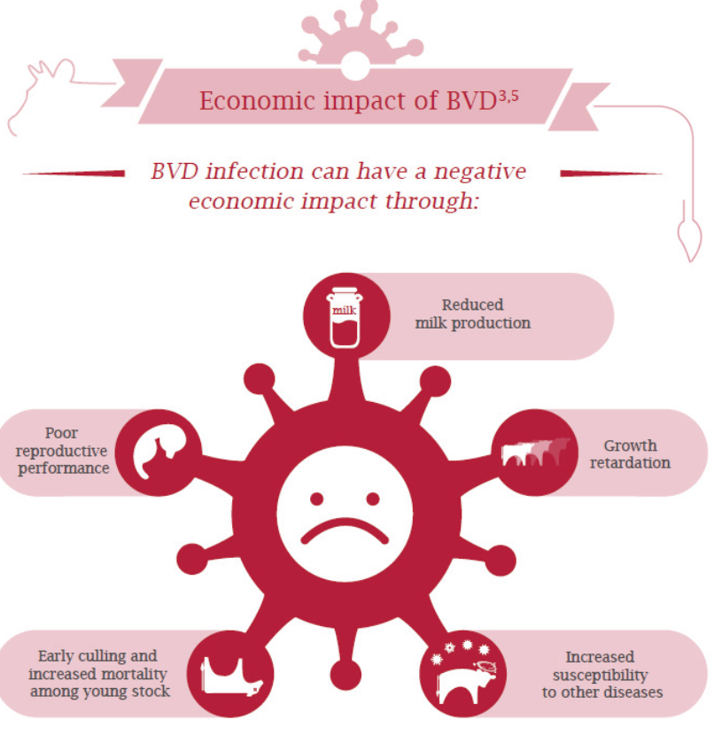
Liver Fluke (Fasciolosis) is a disease caused by the flatworm Fasciola hepatica. It affects a wide range of animals, including sheep and cattle, and although humans can become infected, although this is very rare in the UK. The disease is estimated to cost the UK livestock industry around £300 million every year due to liver condemnations, animal deaths, and reduced productivity.

WHERE DOES IT COME FROM?

The liver fluke life cycle begins when eggs are passed onto pasture in animal faeces. These hatch and infect a small mud snail (Galba truncatula), where they multiply and develop. Under suitable wet conditions, immature fluke called cercariae leave the snail and encyst on vegetation as metacercariae — the highly infectious stage.

Animals become infected when they graze contaminated pasture. The ingested metacercariae develop into juvenile flukes that migrate through the liver, causing significant damage, before moving into the bile ducts where they mature into adults. Adult fluke produce eggs that pass out through the digestive tract and continue the cycle.

It takes approximately 10–12 weeks from ingestion of metacercariae for adult fluke to be present in the bile duct.

Unlike gut worms, animals do not develop immunity to liver fluke, meaning all ages are susceptible.

CLINICAL SIGNS

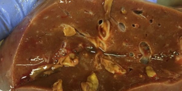
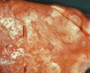
Disease is caused both by juvenile flukes migrating through liver tissue and by adult flukes feeding in the bile ducts.

SHEEP
Acute fasciolosis
Caused by large numbers of migrating juvenile fluke. Can lead to sudden death from internal bleeding or secondary clostridial infections (Black Disease). Signs may include extreme lethargy, abdominal pain, reluctance to move, and collapse. Most common in late summer and autumn.
Sub-acute fasciolosis
Fewer migrating fluke cause rapid weight loss, poor fleece quality, inappetence, weakness and depression. Death can occur without treatment. Typically seen in autumn.
Chronic fasciolosis
Caused by adult fluke in the bile ducts. More common in late winter and early spring. Signs include poor body condition, anaemia and bottle jaw (fluid swelling). Results from impaired liver function and inflammation.

CATTLE
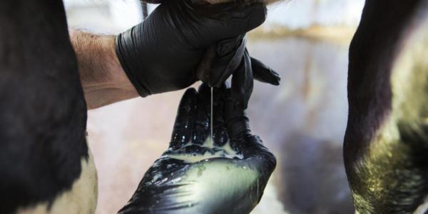
Cattle typically develop chronic disease, shown by poor weight gain and anaemia. Sub-clinical infection can reduce milk yield. Diarrhoea is possible but usually linked to secondary Salmonella infection.

DIAGNOSIS – PREVENTION – TREATMENT

Diagnosis

Liver fluke can be detected through bulk milk, blood tests, or faecal testing. Different tests identify infection at different stages, so test choice depends on the time of year and level of risk.

Prevention

Fluke risk varies greatly between farms and even between fields. Wet, boggy or slow-draining areas are high-risk grazing zones. Identifying and, if possible, fencing off or avoiding known “flukey” areas during peak risk periods can reduce infection.
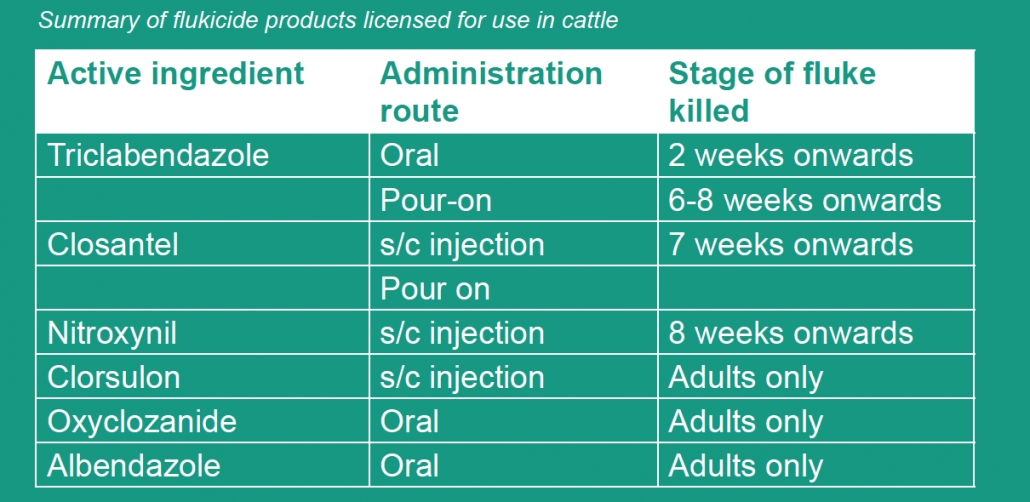
Treatment

Choosing the correct treatment depends on the age and stage of the fluke present, as not all products are effective against every stage. Understanding seasonal risk and test results is essential when planning treatments.

Please get in touch for any information regarding fluke and its treatment.

REDUCING THE IMPACT OF E. COLI MASTITIS ON YOUR HERD

REDUCING THE IMPACT OF E. COLI MASTITIS ON YOUR HERD

Mastitis remains one of the most expensive diseases in dairy cattle, typically costing £250–£300 per case. E. coli mastitis has been shown to reduce milk production by around 15%, equating to approximately 200 litres per cow per lactation, Clinical signs range from mild changes in milk to rapidly developing toxic mastitis which all to commonly can result in death.

E. coli is generally considered an environmental pathogen with infection resulting from bacteria in the cow’s environment gaining access to the udder. E. coli is commonly found in the gut of healthy cows and passed into the cow’s environment in faeces. In order to cause mastitis, E. coli must first gain access to the teat end then migrate up the teat canal and finally become established in the udder.

REDUCING THE CHANCES OF E.COLI

Bedding choice significantly influences the ability of the bacteria to multiply, with sand being lowest risk. Whilst sand beds still need to be maintained it is much more difficult for bacteria to multiply in it. Organic materials such as sawdust or straw can work well but it’s vital that they are clean and dry before applying, moisture is important for bacteria to multiply.

Bedding conditioners such as lime have been found to reduce E. coli’s ability to multiply by drying the bed and raising the pH, but the action is short lived; 24-48 hours. So generally, they need to be applied daily.

Dietary issues that cause loose faeces lead to dirtier udders. Rapid ration changes and SARA are common contributors. Vitamin E and selenium deficiencies weaken immunity and increase severity.

Historically we’ve often seen a higher incidence of severe toxic cases in robotic milking systems. Newer designs and management strategies have gone a long way to reduce the risks. The increased risk is thought to be caused by a number of factors including, the potential for irregular milking intervals in some cows, the ability of the robot to deal with more severe teat contamination during the premilking prep and footbathing immediately exiting the robot splashing teats. Generally, most of these issues can be overcome with changes to robot setup or routine.

There is a commercial vaccine available in the UK, Startvac® which has been shown to reduce severity of E. coli, mastitis. Though it does not reduce the number of cases., it is a useful tool in herds struggling with severe cases as significantly more of the cases only present with milk changes.

TREATMENT

Rapid treatment is vital as mild cases can quickly become toxic.

Recommended approach:

Anti-inflammatories (NSAIDs) and fluids are the mainstays of therapy. All cases should receive an NSAID such as Metacam or Ketofen to reduce inflammation and oral fluids. Severe cases often benefit from intravenous fluids, traditionally reserved for recumbent cases, we’ve seen improved responses from dripping standing cows with severe E. coli. Oxytocin can help with milking out, reducing the amount of toxin the cows is exposed to. Frequently the cows are also low in calcium and so supplementation may be beneficial. Antibiotic use in these cases remains controversial but is generally indicated in more severe cases as bacteria can be found in the blood of up to 30% of cases.

CONCLUSION

E. coli mastitis is common across dairy farms, but risk can be significantly reduced through clean bedding, good hygiene, nutritional stability, and proactive management. Early treatment greatly improves outcomes, and vaccination can help reduce severity on affected farms.

Sam’s trip to a bovine transfer unit in Canada

This month Oakhill Farm Vet Sam travelled across the Atlantic to Alberta, Canada to visit a bovine embryo transfer unit.

Sam had an interesting insight into the bovine industry there…

[checklist icon=”fa-arrow-circle-right” iconcolor=”#39aa87″ circle=”yes”]

  • The main industries in Alberta are oil and beef.
  • Alberta has the second largest total farm area in Canada.
  • Alberta’s feeder cattle (steers and heifers for feeding or slaughter) account for 59.6% of the national total and the beef breeding stock accounts for 42.3%.
  • The predominant breeds that were dealt with at the embryo transfer unit were Angus, Charolais, Hereford and Simmental.  
  • Typical herd size was 200-300 cows.
  • Artificial insemination is common practice in the beef industry.

[/checklist]

The climate is very challenging in the winter. Temperatures can plummet to -20°C to -30°C in January when pedigree herds are calving! Keeping water troughs thawed out and keeping on top of frost bite damage to calves’ ears were particular challenges. The region consists of very marginal grazing land with some barley grown. In mid-may the grass had only just started to think about growing, whilst back home many 1st cut silages were over and done with!

Cattle handling facilities were excellent; hydraulic squeeze crushes were the norm in the region allowing for safe and efficient handling. The cows were of a moderate size with excellent maternal characteristics. Calving ease was a very important trait selected for by farmers. Calves with a birthweight over 100lbs (45kgs) were not retained for breeding irrespective of pedigree. Meat quality is important to the Canadians; marbling is a very desirable feature.

Due to geographical constraints, the majority of cattle travelled to the ET unit for flushing or implanting. The methods and materials used to perform embryo transfer were very similar to here in the UK.

Frozen embryos were shipped all over the world: UK, USA, Argentina, Australia, Thailand to name just a few countries. Introducing genetics into a country through use of embryo transfer is very useful in terms of infectious disease control.

For further information on embryo transfer, please contact the Oakhill Farm Team.

Mobility Scoring

Early identification and prompt treatment is one of the cornerstones of lameness control within the Healthy Feet Programme. Numerous studies have confirmed the benefits of early treatment and we would all accept that treating a lame cow sooner rather than later can only be a good thing.

However, treatment of these early onset cases of lameness is often delayed as other cows get prioritised at the foot trimming visit. For instance, chronic score 3 cows, acute score 3 cases and dry off trims generally make up the bulk of the cows presented to the foot trimmer, rather than the score 2 cows.

Early identification and prompt treatment often requires no extra capital investment and can make a huge difference to the number of lame cows in as little as 6 months.

There are also significant long term benefits as lameness leads to chronic bony changes in the hoof, which in turn predisposes cows to repeat lameness events in subsequent lactations.

Mobility scoring has often been seen as an unwelcome paperwork exercise imposed by outside agencies. On the other hand, we feel that monthly mobility scoring to generate treatment lists is extremely beneficial and can be a very cost effective way to tackle lameness in your herd.

We offer monthly mobility scoring through our Vet Tech service.

These visits often coincide with milking and the data is recorded through our on-farm software App.

We can then generate treatment lists for you or your foot trimmer. In addition, we can also add the results from the foot trimming records back into the software.

This allows us to monitor lameness levels, lesion prevalence and treatment success over time.

For more information about our Vet Tech Service, please call 01772 861300

Sheep – The Big Five diseases

The Big Five Diseases in Sheep

Maedi Visna

Maedi Visna - sheep diseaseMaedi Visna is a wasting disease causing chronic pneumonia. It is caused by a highly contagious virus. Most sheep are infected as lambs via colostrum or aerosol, however generally do not show signs until they are 4 years old.

The main signs are:

[checklist icon=”fa-arrow-circle-right” iconcolor=”#39aa87″ circle=”yes”]

  • Thin ewes/wasting away
  • Pneumonia
  • Mastitis
  • Arthritis
  • Reduced fertility
  • Smaller weaker lambs which grow poorly due to a lack of milk

[/checklist]

Blood tests are commonly used to diagnose MV. Selecting thin cull ewes to test will help identify the disease if present within the flock.

If you suspect MV in your flock or wish to know your MV status we can do a 12 cull ewe screen.
Please call 01772 861300 to discuss.


Ovine Pulmonary Adenocarcinoma – OPA

Ovine Pulmonary Adenocarcinoma - OPA - sheep diseaseOvine Pulmonary Adenocarcinoma is caused by a virus leading to a progressive and fatal infectious lung cancer of sheep. The virus is spread from infected sheep by aerosol and nasal discharge. It is often seen in older thin sheep. There is no treatment.

The signs are very similar to chronic pneumonia:

[checklist icon=”fa-chevron-circle-right” iconcolor=”#39aa87″ circle=”yes”]

  • Difficulty breathing
  • Weight loss
  • Excessive watery fluid from the nose
  • Sudden death
  • Secondary infections such as Pasteurella are common.

[/checklist]

Once signs are evident the disease is fatal.

The definitive diagnosis is made by post mortem examination of the lungs.

If there is suspected OPA ultrasound examination of the lungs can be considered.


Johne’s Disease

Johne's disease in sheep
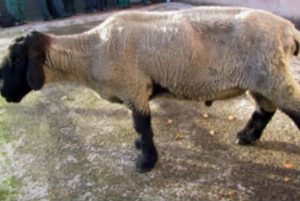
Johne’s disease is caused by a bacterium which grows very slowly and lives for a long time in the environment. It is mainly spread through faecal contamination of feed and the environment, ewes also pass it to their lambs across the placenta and in colostrum. It is the same agent that causes Johne’s disease in cattle.

Infection generally occurs early in life, sheep often do not show signs for years and act as carriers, these are referred to as ‘sub-clinically infected’.

Affected sheep suffer from:

[checklist icon=”fa-chevron-circle-right” iconcolor=”#39aa87″ circle=”yes”]

  • Severe weight loss
  • Sudden death
  • Anaemia
  • High parasite burdens
  • Affected flocks suffer from poorer production, more thin ewes and increased culling.

[/checklist]

Testing for Johne’s can be tricky at an individual level. There is no single diagnostic test that can detect the disease at all the stages and sub-clinically infected animals may test negative.

Blood screening groups of cull/thin ewes can be effective at identifying Johne’s disease in your flock.


Border Disease

border disease in sheep
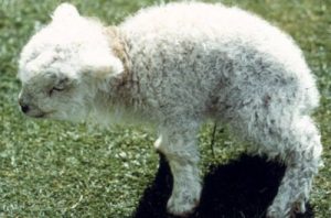
Border disease or hairy shaker disease is a virus which causes birth defects, barren ewes and abortion. It is from the same family of viruses as BVD in cattle.

If a ewe is infected during pregnancy the virus passes through the placenta to the lamb, in some cases this causes embryonic loss or abortion. Some lambs survive and are born Persistently infected (PI). PIs shed the virus continuously and can infect other sheep and lambs. This can be a major problem in naïve flocks unknowingly buying in PIs which can then cause around half of lambs born to be affected by the disease.

[checklist icon=”fa-chevron-circle-right” iconcolor=”#39aa87″ circle=”yes”]

  • Infection in non-pregnant adult sheep is generally short lived and mild, often no clinical signs are seen.
  • Hairy shakers are lambs infected with the virus whilst developing in-utero effecting their nervous system which leads to trembling and incoordination. They also have long curly wool.
  • Some lambs are born completely normal but maintain the infection, these are called Persistently Infected animals (PI).

[/checklist]

Hairy shakers are usually a good way to diagnose the presence of the disease. These lambs can also be blood tested to confirm presence of the virus.

Blood testing a group of ewes can identify if the flock has been exposed to the disease and is likely to be present.


Caseous Lymphadenitis – CLA

Caseous Lymphadenitis - CLA - sheep disease
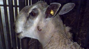
CLA is a chronic bacterial infection of the lymph nodes resulting in abscesses. The bacteria often enters via cuts and is commonly spread through close contact when animals are housed or are trough fed. The disease can also be transmitted indirectly such as on shearing equipment.

[checklist icon=”fa-chevron-circle-right” iconcolor=”#39aa87″ circle=”yes”]

  • Infected animals have lumps often behind the jaw, in the arm pit and in the groin. These lumps are often cold and painless but can burst open and drain pus. Once they have healed and scarred over the abscess can re-occur.
  • Abscesses also form inside the sheep causing ill-thrift and weight loss.

[/checklist]

Lumps and abscesses are a classic sign of the disease and should not be ignored.

Diagnosis is either by sampling the intact abscesses and culturing the pus or by blood test.


If you would like further information, please contact our Farm Team.

Neonatal Lamb Care

Neonatal Lamb CareLambing time is the most crucial part of the year for making your sheep business profitable. Lamb deaths from birth to 3 days old should be less than 7% however many farms range between 10- 25%.

A major factor influencing mortality in neonate (young) lambs is ewe body condition; as this directly relates to lamb body weight, colostrum quality and quantity. It is therefore important to ensure ewes are at BCS 2.5 to 3.0, (so vertebral spinous processes only felt with pressure and transverse processes only felt with hard pressure), and they are on a rising plane of nutrition in the run up to lambing. Energy and protein blood profiles can be taken 2 to 3 weeks before lambing to ensure ewes are receiving the correct nutrition to help reduce cases of twin lamb disease.

Another major cause of neonatal mortality is watery mouth. These lambs typically are lethargic, salivate and have a distended abdomen. It is caused by Ecoli and picked up by ingestion. Treatment involves electrolytes and antibiotics but is often hopeless. Therefore the emphasis should be on prevention which involves keeping pens clean, (especially towards the end of the season), dipping navels with strong iodine and ensuring all lambs get enough good quality colostrum quickly. See below:


Always ensure colostrum is…

QUALITY

Ensure ewes in good BCS, if you’re uncertain as to the quality of colostrum; colostrum quality can be measured using a refractometer: aim for specific gravity >1.05. Only use sheep colostrum replacement (colostrum from a milking sheep farm will usually be better quality than powdered colostrum); cow colostrum can cause a fatal anaemia. Also remember to give your Clostridial and Pasteurella vaccine four to six weeks before lambing to ensure the lambs receive antibodies against these diseases. (Any ewes that haven’t had this vaccine before will need a course of two injections four to six weeks apart).


QUANTITY

200ml/kg within 18 hours of birth, with a maximum of 50ml/kg intake on each occasion.

Remember lambs in adverse weather conditions will require more colostrum or milk as more energy from their feed will be used to keep them warm.


QUICKLY

Within 2 hours of birth.


SQUEAKY CLEAN

Ensure ewe udders are clean and dry and the equipment to mix and administer colostrum is cleaned and disinfected between lambs.


QUANTIFY

We can also look if your lambs have received enough colostrum by blood testing any less than a week old.


Finally, there is continual pressure on antibiotic use. So to ensure we don’t end up with a world full of super bugs we need to be careful how we use antibiotics. Preventative use of antibiotics, such as blanket use of Spectam or Betamox LA for watery mouth needs to be reduced as this will actively select for resistance and there is pressure from Red Tractor Assurance to reduce this practice.

We recommend only using antibiotics….

    • Towards the end of the lambing season, when the sheds have a build up of Ecoli.
    • In triplets, when their ewe may have poorer quality and quantity of colostrum
  • In lambs from ewes in poorer condition, as the quality and quantity of colostrum will be poorer.

Also remember entropion, where the lamb’s eyelid is inverted causing the eye to be held shut, tear staining and damage to the eye. This is a heritable condition so affected lambs should not be kept as replacements, ewes should not be used to breed replacements and consider ram choice if a large percent of flock are affected.

If you have any questions regarding lamb care, please contact the Oakhill Farm Team.

Mastitis in ewes

Mastitis in ewes can be fatal and usually results in the end of her productive life.

The usual presentation is acute gangrenous mastitis (blue or black bag) usually caused by either Staphylococcus Aureus or Mannheimia Haemolytica. S. Aureus is normally present on the teat skin but damage to the teat ends allows it access into the teat canal and causes mastitis.

M. Haemolytica is present in the tonsils of lambs so colonises the udder during suckling. The bacteria produces toxins which prevent blood flow and cause the udder to turn blue/black hence the name. This often occurs around peak lactation (three to six weeks post-lambing) and the initial clinical signs may be as subtle as a ewe looking lame or a lamb bleating because it’s hungry. The disease then rapidly progresses.


The risk factors include:

[checklist icon=”fa-arrow-circle-right” iconcolor=”#39aa87″ circle=”yes”]

  • Under feeding ewes round lambing or ewes in poor condition, as lambs to these ewes will cross suckle or create teat lesions by over suckling.
  • Damaged teat end or Orf on teats
  • Lumps in the udder
  • Lambing inside (the longer the time spent inside the greater the risk of developing mastitis)
  • Dirty, wet pens and dirty hands when handling udder/ underneath of sheep
  • Harsh, cold weather

[/checklist]


Treatment is unlikely to save the udder but may save the ewe’s life:

[checklist icon=”fa-arrow-circle-right” iconcolor=”#39aa87″ circle=”yes”]

  • Remove lambs from the ewe and ideally artificially rear as rearing these lambs on another ewe may spread the mastitis
  • Antibiotic injectable AND non-steroidal anti-inflammatory such as Loxicom
  • A ewe that has had mastitis will be more likely to get fly strike so preventative fly treatment is essential.
  • There is a vaccine against S. Aureus available; VIMCO. This has to be given three and five weeks pre-lambing. Most of the trial work has been done in dairy ewes and it is licenced to reduce to the incidence of subclinical mastitis. Potentially this could also help to reduce the incidence and severity of black bag in your flock however at the moment there is limited evidence to prove this.

[/checklist]

If you have any questions regarding mastitis in your flock, please contact our Farm Vet Team.

Extended liver fluke season and climate change

Fascioliasis

An important stage of the Liver fluke life cycle takes place in mud snails and requires wet and warm conditions (above 7 – 10°C) during the summer months. Over the last couple of years unseasonal weather has seen these conditions well into autumn and winter. This means that sheep and cattle are still being infected later in the season and more frequent testing and treatment may be required.

Fascioliasis can have a serious financial impact on a sheep farm with immediate losses up to 10 per cent caused by acute/subacute disease. Chronic disease could half profits by reducing lamb crop and increasing ewe mortality. Lack of treatment in cattle will lead to reduced growth and yields and discarded milk if treatment is no targeted.

We recommend testing your sheep and cattle for the presence of Liver fluke. Treatment needs to be targeted at your flock/herd and will vary from farm to farm. Please discuss your individual requirements with your vet to develop a treatment plan for the coming months.

liver fluke life cycle

BVD -STAMP IT OUT

A new initiative has been launched to help farmers in England tackle BVD (Bovine Viral Diarrhoea).

It is currently estimated that BVD costs British farmers up to £60million/year, reducing productivity and profitability as a result of poor growth rates, poor health with increased pneumonia and diarrhoea, reduced fertility with higher rates of foetal death. Rigorous testing of the herd is vital to help eradicate the disease from the unit.

BVD FACT
The estimated cost of BVD is between £13 and £31 per cow in Great Britain.

The £5.7 million funding package from the Rural Development Programme for England (RDPE) is managed by SAC consulting and is the largest BVD project launched in England. Farmers will be able to apply for free on farm health planning for BVD control. Keepers of breeding cattle will be eligible for the scheme, which will help to assess the BVD status of the herd and develop a control plan.

After attending the first meeting a one to one on farm visit will be undertaken to carry out a check test on the herd. If the results indicate active infection then a follow up visit will be arranged to develop a customised action plan which may include further testing (PI hunt). The overall aim will be to reduce, eliminate or prevent BVD within the herd.

Beef & Youngstock – Housing Considerations

We have experienced a drier summer than we are used to and this has posed different challenges. Worm burdens have been greatly reduced on pasture over the summer however if we get a heavy rainfall following this dry period there is likely to be mass emergence of worms onto pasture which may have severe consequences for our livestock if left untreated.

Exposure to lungworm may also have been reduced over the summer and so we would advise watching out for or listening to cattle to see if they are coughing over the next few months and treating if necessary. Lungworm can cause significant respiratory disease and death in cattle and should not be overlooked in any grazed stock. Where cattle have been grazed for extended periods the use of long acting anthelmintic bolus activity may have worn off and further treatment may be necessary.

Testing for Lungworm larvae can be done on a faecal sample however is slightly different to a normal worm egg count. If you would like to check grazed stock for Lungworm please submit a faecal sample stating that you require Lungworm testing.

Bringing in cattle from markets, other farms or common grazing can introduce new diseases to your own farm, including parasites such as liver fluke. Liver fluke is a common parasite that infects cattle, sheep and other domestic animals as well as wildlife such as deer and rabbits. Following several wet years monitoring, testing and treating for liver fluke early is also recommended during this year.

Resistance of liver fluke to treatments, in particular products containing triclabendazole (TCBZ), appears to be an increasing problem and one that needs managing on all livestock farms. It is important that care is taken to reduce selection pressure for resistance whenever possible by only using triclabendazole-containing products when no other options are suitable. Other medicines which contain the active ingredients, closantel, clorsulon, nitroxynil, oxyclozanide and albendazole are effective against adult flukes and some (nitroxynil and closantel) are effective against late immature stages between seven and eight weeks old.

If treating now or immediately on housing we would recommend using a Triclabendazole product but if cattle are housed for 7 weeks or more, one of the other products is often more suitable. Please speak to us about testing or when to treat.