Call Us
Goosnargh:
Lytham Road:
Kirkham:

MRI provides the key to the diagnosis for Toby

Toby, a 7 year-old, Cob cross pony presented to Oakhill with a several week history of mild, right forelimb lameness. Despite a short period of box rest and pain-relief, the lameness did not resolve, and a veterinary opinion was sought.

Nikki Platt, our senior lameness veterinary surgeon examined the pony and noted that the pony’s front feet pointed in slightly, and that the outer walls of his hooves were slightly longer than the inner walls. When observed moving, Toby was sound in a straight line, 3/10 right forelimb lame on the left rein and 2/10 right forelimb lame on the right rein. The lameness was slightly more obvious on a firm surface. The pony did not resent standing with his limbs flexed up, and this procedure (known as a flexion test) did not cause any increase in lameness.

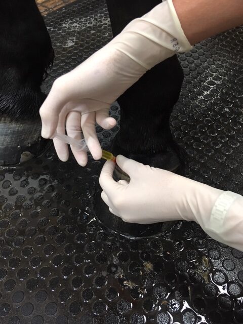
In order to be certain of the origin of the lameness, diagnostic analgesia was performed (where local anaesthetic is used to remove pain sensation from an area). A palmar digital nerve block was used to de-sensitise the right foot. This caused the resolution of the right forelimb lameness, and the appearance of a mild left forelimb lameness was present when the horse was lunged. It is common when performing diagnostic analgesia that, having eliminated the most significant pain from the lame limb, the horse then begins to show lameness in the opposite limb, as the horse is a symmetrical animal after all!

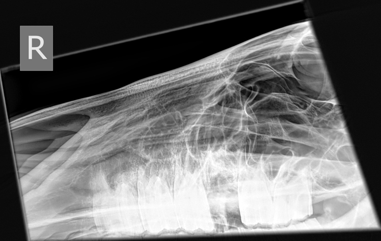
Having conclusively identified the source of pain, x-rays of the feet were taken. In this case, the bony structures of the foot were unremarkable other than to identify a slight compression of the coffin joint space on the medial (inside) aspect compared to the lateral (outside). The lack of explanation for the cause of the lameness on the radiographs meant a need to assess the soft tissues of the foot by performing MRI (magnetic resonance imaging).

The procedure was carried out under mild sedation with the pony standing. Following the evaluation of the 400+ images of Toby’s feet we obtained, the diagnosis was clear – moderately severe collateral ligament desmitis (inflammation) of the coffin joint in both front feet. 

The collateral ligaments are responsible for stabilising the movement of a joint. If (like with this pony’s ‘toe in’ conformation) there is a slight twist in the limb, a joint can be loaded unevenly across its surface during weight bearing. This means that one or both of the ligaments may be subject to more strain than it is designed to take whilst stabilising joint movement. This repetitive, excessive strain leads to microscopic damage of fibres within the ligament, causing inflammation, pain and scarring.

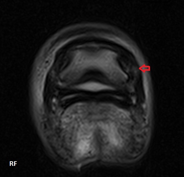
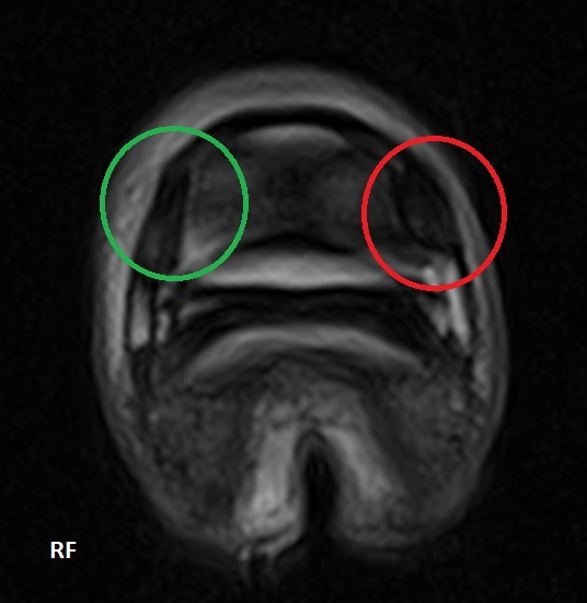
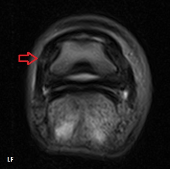
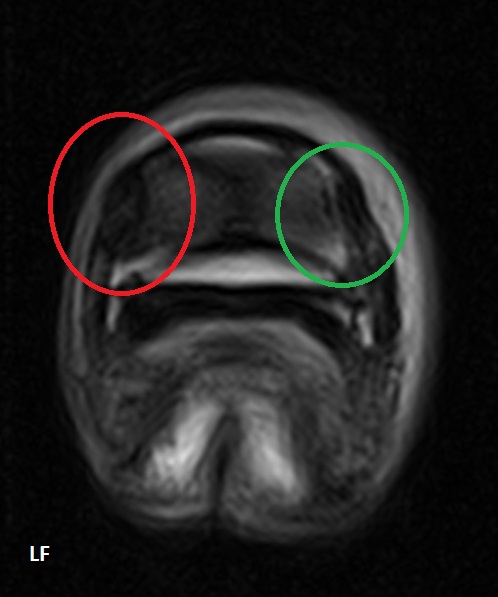
On the MR images pictured, the red arrows and circles indicate the damaged medial collateral ligament (with the green circles highlighting the comparatively normal lateral ligament). The damaged ligaments have an ‘increased signal intensity’ i.e. they have a brighter, whiter appearance, due to the infiltration of inflammatory fluid into the structures. The damaged ligaments are also larger, partly due to this fluid infiltration, and partly due to scar tissue being laid down.

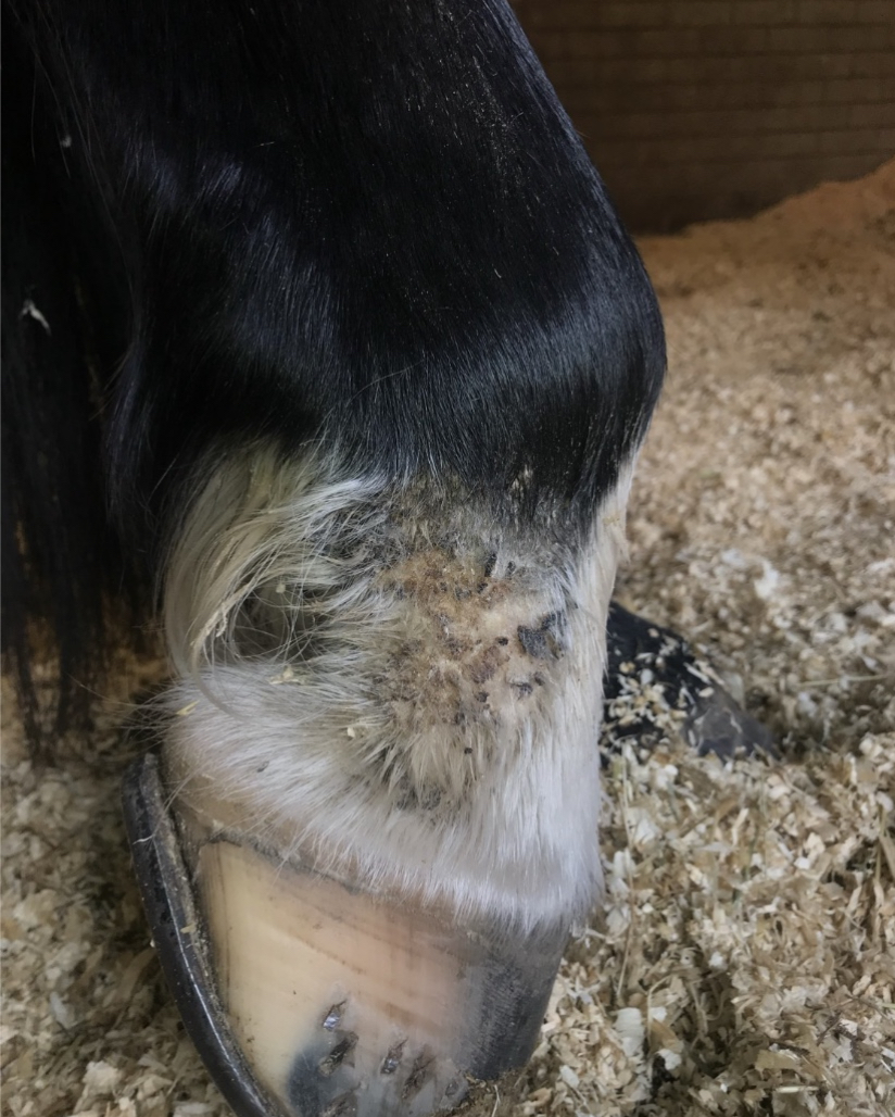
MRI was crucial in this case to reach a diagnosis. In reaching a diagnosis, we were able to advise on appropriate treatment. In the first instance, this horse was prescribed six weeks of box rest with a gradually increasing walk exercise programme. The farrier’s involvement was also crucial in this case – the pony was trimmed to maximise the symmetry of the foot (the farrier was able to work from the radiographs provided) and shod in heart bar shoes to help to stabilise the way it was landing. 

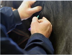
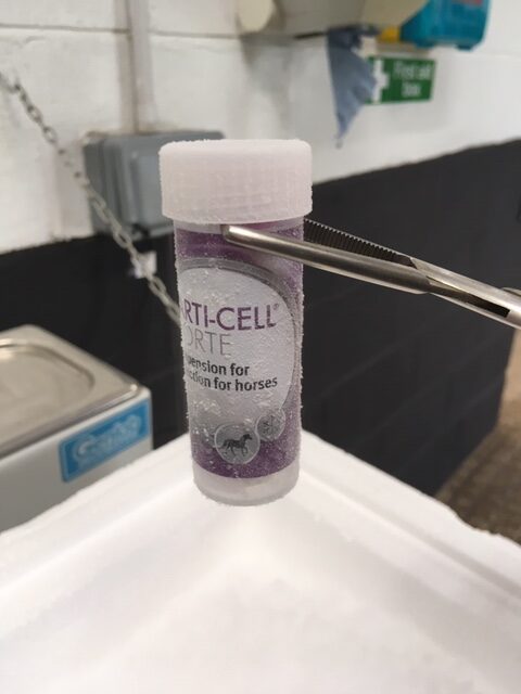
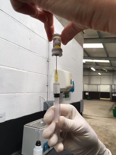
After six weeks, the pony was greatly improved, showing just 1/10 right forelimb lameness on the left rein only. Having given the soft tissues adequate time to rest, a low dose of steroid was injected directly into the coffin joint, to resolve the mild inflammation that remained at the margin of the ligaments. One month later, the pony was sound!

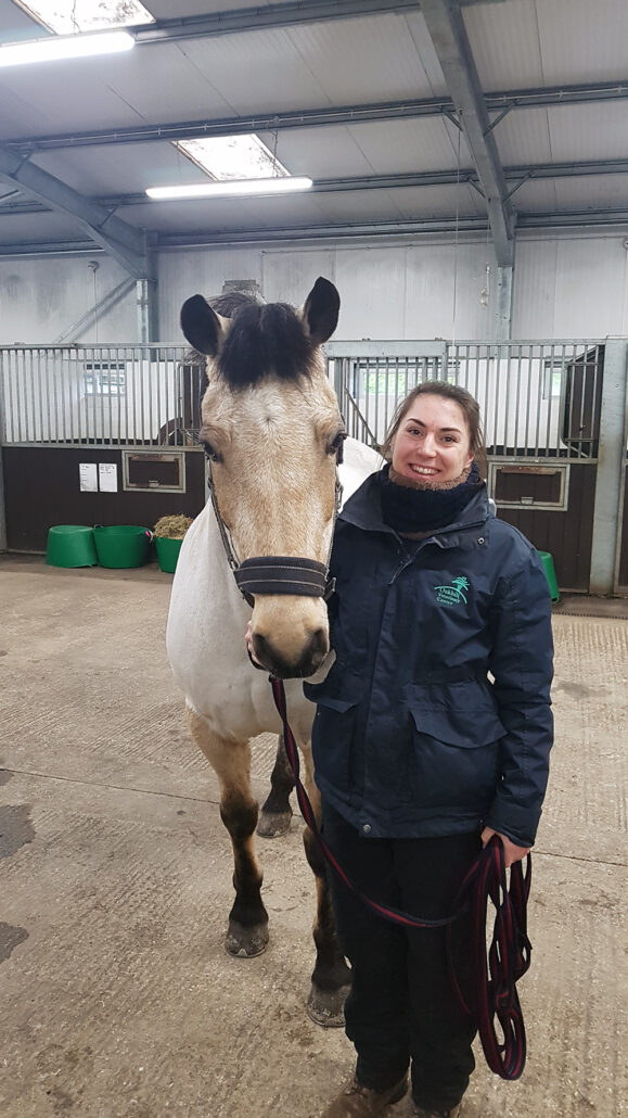
Toby is now successfully building back up to his previous level of exercise, and is anticipated to remain sound with ongoing maintenance of good foot balance.

Why is my horse’s sheath swelling and how can I tackle the winter worm burden in my horse?

A tumourous growth on the end of
this horse’s penis

To continue our series on Winter medical conditions, in this newsletter, we are going to cover swollen sheaths and worming your horse at this time of year.

Swollen Sheaths

It is quite common for geldings/stallions to present to us with swollen sheaths over the Winter months. More often than not, sheath swelling results from the affected patient being stood in for prolonged periods of time which sadly, is inevitable with deteriorating weather conditions. Swelling should improve, if not resolve, with exercise/increased movement.

Other causes of swelling include excessive accumulation of smegma, low blood protein, infection and fortunately less commonly, infection secondary to penile tumours (squamous cell carcinomas). Fat can also accumulate in the sheath but this has a more gradual onset.

Extensive cancerous
(squamous cell carcinoma)

Should excessive smegma occur, then cleaning the sheath and penis is indicated. Sheaths should not be over-cleaned as this can disrupt the normal flora (bugs) that should be present to maintain ‘normal’ sheath health. 

Low blood protein can result from small encysted redworm and colitis to name but two potential causes. Diagnosis can be suspected based on history and compatible clinical signs but blood test results are confirmatory.

With infectious causes of sheath swelling, the sheath is firm, hot and painful to palpate. Your veterinary surgeon will examine and clean the sheath and penis under sedation in addition to prescribing antibiotic and anti-inflammatory drug therapy. 

Plaques – an earlier cancerous change

Penile tumours sadly occur but fortunately are not terribly common. The tumorous growths vary in appearance from small white plaques to large proliferative growths.

Treatment options depend on the stage of the disease at presentation. Surgical removal is indicated, if possible.

Elevate your vaping experience with Lost Mary Vape’s stylish innovation. Discover more at http://lostmaryecig.co.uk/ and redefine your ritual.

Worming

A common question we are asked at this time of year is ‘what wormer, if any, should I use for my horse?’

https://www.high-endrolex.com/11

At this time of year, we should cover horses for small encysted red worm (cyathostomes). Suitable wormers include a single dose of moxidectin (which is the drug found in Equest and Equest Pramox) or a 5-day course of fenbendazole (Panacur Equine Guard).

Sadly, due to overuse of wormers in the past, a huge amount of resistance to fenbendazole exists. This highlights the importance of practising responsible worming and seeking your vet’s advice to devise a suitable control programme for your horse.

A blood test for small encysted redworm has recently been developed. This means that we now have diagnostic tests available for roundworm, tapeworm and small encysted redworm. Testing for small encysted redworm should be performed between September and April. The test is not suitable for horses who have had high faecal worm egg counts throughout the rest of the year; these horses should be covered with a suitable wormer, as per above, regardless.

Keep your eyes peeled over the next 4-6 weeks for the launch of our 2021-2022 Equine Worm Control Plan; more information to follow! As always, if you have any questions on worming, please contact our team. 

Is my horse’s poor ridden performance due to discomfort?

We’ve all been there! Feeling frustrated that your horse won’t pick up the correct canter lead, or does he/she disunite behind in canter? Will your horse perform lateral work happily one way and not the other way? Does it struggle to use it’s hindlimbs correctly to provide power to the gait? Or does it have an annoying hopping like gait on the bends?! These are all complaints that we are used to investigating every single day. Did you know that we have talented riders amongst our nursing team who will happily ride your horses on our arena whilst we investigate the ridden problem? 

As horse owners, we know that our horses are desperate to please us most of the time! Don’t get us wrong, there is the odd occasion when they push their luck of course with a little bit of cheekiness, but on the whole they want to do a great job for us as their riders. So when they are objecting to what we are asking them to do, we need to stop and ask these questions: 

  • Is my horse at a suitable age to be able to do what I am asking? 
  • Is my horse adequately trained and prepared for what I am asking them to do? 
  • Are there any external factors or management changes that could be impacting my horse’s ridden behaviour? 
  • Am I asking something that is out with the athletic capabilities of my horse? 
  • Could my horse be in discomfort? 

Sometimes, by simply examining the musculoskeletal status of your horse, we can help you to make adjustments to your feeding or training regime to help strengthen the weaker areas of the horse. Further investigation is not always warranted or recommended!

As highly trained professionals, we are here to advise you regarding the best way to proceed with your horse, that may be a bute trial, a lameness investigation, x-rays of the spine or gastroscopy……Horses are unique, and the way they present is unique, you are unlikely to find a solution to the problem on Facebook 🙂 and are often much better bringing your horse to our clinic for an initial assessment with one of our experienced veterinary surgeons in this field.

We are more than happy to begin a discussion with you by phone if you have any concerns about your horse’s ridden performance, we are always here to help.

Equine Vaccine update

New year, a fresh start. What better time to check our horses vaccinations and ensure they are up to date. Vaccination seems to be the word of 2021 but sometimes the information surrounding them can be overwhelming. Vaccines are important not only for preventing diseases but also for slowing disease transmission and reducing the severity of clinical signs.

Horses that compete or attend organized events require 6 monthly flu boosters. Vaccination amnesties were put in place in 2020 due to COVID 19. However, the major governing bodies have either since returned to 6 monthly boosters or are yet to announce 2021 regulations. We recommend to keep up to date with annual vaccines and when competitions return boosters can be administered if needed. Remember to always check with the organizers about their specific rules.

In the UK, horses and donkeys can be vaccinated against various diseases but the most common conditions to vaccinate for include Equine Influenza and Tetanus.  With reports of sporadic cases of equine flu in the north west area and a case of tetanus recently treated by one of our vets, here is a brief reminder of these diseases and what to look out for. 

Equine Influenza is endemic in the UK, it is a highly infectious virus that affects the respiratory system. It spreads rapidly among equine populations and can be passed on via direct contact or a contaminated environment. Equine flu can be a very debilitating disease, requiring a long recovery period and in rare cases can even be fatal.  Signs to look out for include: Fever, harsh dry cough, nasal discharge, loss of appetite and depression.

Tetanus is a dangerous and often fatal disease that can affect any horse or pony.  Clostridium tetani bacteria found in the soil can contaminate even a minor wound. The bacteria produce a toxin that causes muscle spasms, paralysis and death. Every horse or pony should be vaccinated for tetanus. 

If you are vaccinating for the first time or the vaccines are out of date, here is a reminder of the vaccine schedule: These vaccines can be administered in combination or separately.

Equine Influenza

  • 1st vaccination – Foals can start from 6 months of age
  • 2nd vaccination – 4 weeks later  (21- 92 days after the 1st)
  • 3rd vaccination – 5 months later (150 – 215 days after the 2nd)
  • Annual booster – within 365 days of the last vaccine. 

Tetanus

  • 1st vaccination – Foals can start from 6 months of age
  • 2nd  vaccination – 4 weeks after the 1st vaccination
  • 1st  booster – 12 months later
  • Subsequent boosters administered every 2 years

Other diseases that horses can be vaccinated for include Equine Herpes Virus, Equine Viral Arteritis and Rota Virus. Our Equine Care Plan’s make it easy and affordable to keep up to date with vaccinations.  Please call or send us an email if you have any questions regarding vaccinating your horse.

Heather Stephenson Bio

Growing up in Manchester I spent all my free time at my local riding school, and I was extremely fortunate to have my own horse and supportive patients who gave up their weekends to take me showjumping.

This hobby soon developed into my passion, and after completing a BSc Equine Science (Hons) with Hartpury College, I started my career as an Equine Technician at the Animal Health Trust, Newmarket.

After 5 years down South, I recently moved back home and began my new role at Oakhill Veterinary Centre. Day to day I look after our lovely patients and assist our vets throughout a variety of procedures and surgeries. I especially enjoy all things imaging, with my principle role being in the acquisiton of MRI scans.

I am working towards qualifying as an Equine Veterinary Nurse so spend a lot of my free time studying but when I can, I enjoy keeping fit and spending time with family.

Infundibular caries treatment in the horse – Fillings!!

Bonita is a lovely, 21 year-old Welsh Cross mare whose owners ensure that her teeth are regularly examined and floated. Several years ago it was noted that her 209 and 109 (4th cheek tooth back on the upper right and left side) suffered from a condition known as infundibular caries. There are two infundibula in the middle of each upper cheek tooth, and consist of an enamel cup which should be filled with a material called cementum. In Bonita’s case this cementum was absent and food and bacteria had caused decay within the infundibula. As the disease progresses the two infundibula can merge leaving a weak point in the middle of the tooth, leaving it at risk of fracture and sequential apical (tooth root) infection.

To preserve the structural integrity of the teeth affected and to preserve the tooth for as long as possible Bonita’s owners decided to restore the infundibula. This involved removing all decayed material and food and filling the cavity remaining with a flowable composite material.

Unfortunately, Bonita’s caries was quite advanced so whilst the filling preserved the tooth for as long as possible (some years) one of the teeth did eventually fracture.

Here is an oroscopic picture of the tooth. As you can see there is a white composite which is the filling material sandwiched between two fragments of tooth. As the fragments have separated, food has travelled up between the two allowing infection to spread to the root of the tooth.

Stuart performed the extraction under standing sedation and a local anaesthetic nerve block which anaesthetises the entire right upper jaw.  Initially the gum is elevated from around the tooth. The gap behind and in front of the affected tooth is then spread using appropriately named ‘spreaders’. Forceps are then placed securely on the tooth and gradual left to right movements are made to stretch the periodontal ligament, which secures the tooth within the socket. When loose enough the tooth is then ‘fulcrumed’ (pulled at a right angle) out from the socket. The socket can then be fully examined and cleaned whilst placing a honey soaked swab to help the socket to heal.

An x-ray taken following tooth extraction shows no remaining root and lots of bony reaction surrounding the socket due to the infection and inflammation that will now subsequently resolve.

Bonita was soon back to eating hay as if nothing had happened. She was discharged home the day after the procedure on antibiotics and pain relief and we are pleased to report that after 4 weeks the socket has completely healed!!!

Horses cope incredibly well with extractions and only usually need 1 week off from ridden work. Bonita’s owners are continuing to keep a close on her for any evidence of further oral discomfort such as quidding and dropping of her feed.

Equine Mites

Mites typically affect the legs of our feathered breeds. Mites can also affect other breeds and one of the more common areas affected in non-feathered breeds is the facial area.

Mite infections can occur throughout the year but as mite populations are highest in Winter, this is the time of year we see the most clinical cases.

Affected horses display signs of itchiness and those with leg mites frequently and repeatedly stamp their feet, bite/chew at their feathers and scratch on various objects to name but a few. On closer examination, crusts & hair loss, with/without weeping lesions are generally present. Some horses markedly object to examination of the lesions so care should be exercised when attempting to examine.

Diagnosis is confirmed by examining a skin brushing under the microscope but frequently diagnosis is based on a compatible history and physical examination findings.

Treatment should ideally start with clipping; however, most owners do not want to proceed down this route in the first instance but in the case of treatment failure, clipping is highly recommended. The limbs should then be bathed in a solution to try soften/break up scabs/crusts prior to the application of a topical treatment.

There are many topical treatments on the market indicating the lack of a gold standard treatment and the fact that different horses respond differently to different treatments; what may work in one patient may not work in another. An injectable treatment is also available. The is a frequently utilised treatment option but it’s use is off-licence in horses.

Following treatment, your horse’s stable should be thoroughly cleaned out to prevent re-infection.

Equine Lice

Moth eaten appearance of a
pony with lice

Our horse’s thicker and longer coats over the winter months becomes an ideal breeding ground for lice, especially when we then cover them in a warm rug. The life cycle of the louse is complete within 4 weeks and eggs hatch 10 days after being laid. Eggs or nits are small (1mm), yellow-white in colour and glued tightly to the base of hairs.

Lice can be seen when this ponies fur was separated

Lice are incredibly contagious and can affect horses of all ages but those kept in large groups indoors, the elderly, young and those with immunosuppressive conditions such as malnourishment and Cushing’s Disease are at an increased risk.

The feeding nature of the lice causes intense skin irritation with horses becoming extremely itchy. This can lead to patchy hair loss and a scurfy coat. In severe infestations the horse or pony can become anaemic.

Lice can be readily spotted by parting the horse’s fur especially along the topline.

Treatment involves topical application of a permethrin or cypermethrin product applied twice at two-week intervals. In contacts should be checked and treated in addition to washing rugs, numnahs, head-collars at a high temperature.

Clipping should also be considered.

Remember, lice are species specific and equine lice do not live on human hair/skin

Asthma/RAO/COPD (and management of):

COPD, also known as RAO and most recently, equine asthma, occurs when inhaled allergens initiate a cycle of bronchospasm (airway constriction) and airway inflammation (increased inflammatory cells and mucous). Two forms commonly occur- Summer asthma secondary to pollen and the more renowned form triggered by dust which can occur throughout the year. It is the latter form which we are beginning to see with increased frequency at this time of year now that our horses are spending more time stabled. 

Affected horses present with clinical sign of varying severity. Some merely have a history of poor performance or a mildly increased respiratory (breathing) rate whereas others are more severely affected and present with respiratory distress. 

Diagnosis is often based on the presence of compatible clinical signs but is confirmed following respiratory tract endoscopy and submission of airway fluid samples for laboratory evaluation. 

With dust-induced asthma, in particular, management changes are of paramount importance. The affected patient should be out, breathing fresh air in so far as possible. Whilst stabled, minimising dust in the patient’s environment is crucial. Utilisation of ‘dust free’ bedding (paper, cardboard, dust extracted shavings) should be considered. Bedding should be laid with the patient outside of the stable with small amounts utilised and changed daily. Cobwebs should be hoovered from the stable environment quarterly. Grooming should also take place outside to minimise stable dust. If feeding hay, investment in a hay steamer should be considered. Otherwise, hay should be thoroughly soaked and fed prior to drying out. Management changes alone in the first instance may not be sufficient and therefore, concurrent drug therapy is frequently indicated.

Drug therapy is based on relieving bronchospasm/airway constriction through the use of bronchodilators and relieving airway inflammation through the use of steroidal anti-inflammatory drugs. Drugs may also be administered to break up airway mucous (mucolytics). Airway inflammation is slow to resolve and treatment may be required for a period of months. In some cases, if ongoing allergen exposure is present, treatment may be required on an ongoing basis or repeatedly when flare-ups occur. Drugs may be administered orally or by the inhalatory/nebulised routes. Your veterinary surgeon will discuss the best method of drug delivery for your horse/pony.

Regenerative therapy

Lameness is a pain-avoidance strategy adopted by horses, and is a common cause of poor athletic performance and compromised welfare.  Whatever the precise cause of pain (e.g. osteoarthritis,  tendon injury), that pain is caused by inflammation. 

Inflammation is the cascade of chemical and cellular events that occurs following any type of tissue damage. By causing pain it alerts the animal to rest the damaged area, thereby preventing further injury. Inflammation also acts as the initial stimulant of the healing or repair process, hence it is extremely important. However ongoing, uncontrolled inflammation causes chronic pain and can actually exacerbate the tissue damage. This is where veterinary intervention becomes necessary.

Conventional therapies aim to stop the inflammatory process, and these remain a vital, cost-effective component of orthopaedic disease treatment.  However, they do not influence the repair of tissue and can occasionally delay this important process. 

‘Regenerative therapies’ aim to optimise the repair of a structure by replacing damaged tissue with tissue of the same cell type and hope to minimise the formation of non-functional scar tissue, hence maintaining the original biomechanical properties of the structure. This increases the probability of return to previous athletic ability, and reduces the likelihood of ongoing lameness and/or reinjury. 

Research and clinical trials of regenerative therapies have been ongoing since 2003, but there are now several exciting options that have been scientifically proven to modify inflammation and reduce pain in clinical trials, all of which are available at Oakhill.

Stem cells are a type of cell which have the potential to develop into a variety of more specialist cell types dependant on the environmental signalling that they are subject to. An embryo begins as a ball of stem cells that go on to develop into every type of cell required to make a mature being! Stem cells continue to be present within the body after birth in reduced quantities.

It is not fully understood how stem cells behave when they are used as a medical treatment – whether they differentiate into the same cell type as the tissue they are introduced to, or if they modulate the inflammatory process. Either way, they have been found to decrease or eliminate lameness when used to treat joint disease, and have the potential to reduce the reinjury rate when used to treat tendon injuries!

Autologous stem cells are collected from the individual horse that requires treatment. Bone marrow is collected (most commonly from the sternum) under sedation and sent away for complex processing to provide a product containing millions of stem cells. 

These cells are injected into core (central) lesions within tendons and ligaments.

Allogenic stem cells are produced from the blood of donor horses (treated to prevent reaction when introduced into the horse requiring treatment). These are purified to get rid of other blood cells and then cultured to increase the number of cells into the millions. They are specifically stimulated to give the ability to differentiate into chondrocytes – the cell type present in cartilage.

Commercially this product is available as Arti-Cell. This has proven highly successful at reducing lameness in horses with degenerative joint disease.

Interleukin-1 Receptor Antagonist Protein, more commonly referred to as IRAP, is a protein synthesised by a variety of cells. It prevents the actions of Interleukin-1 – a substance which has an important role in the induction and maintenance of inflammation within diseased joints. Studies in humans and horses have proved that intra articular IRAP injections reduce synovial (joint lining) inflammation and lameness.

IRAP is produced by collection of blood (from the horse to be treated) in a special syringe.  This is then incubated overnight before filtration to produce concentrated and purified IRAP. This can be frozen to allow storage of the product until an appropriate time for medication of a joint. This product can be of benefit where steroid medication is not appropriate (e.g. competition horses where steroid medication is prohibited, horses at risk of lamintis) or where joint pain no longer responds to steroid medication. There is also some evidence that the effects of IRAP last for up to two years!  

Platelet Rich Plasma, or PRP is simply defined as plasma (the none cellular component of blood) which has been processed to have a high concentration of platelets. It is rich in growth factors – substances which stimulate cell multiplication and tissue repair, therefore it promotes a favourable environment for healing. Like IRAP, it is produced by the specialist collection and processing of blood (from the horse to be treated). This can be done immediately prior to injection of the PRP into the area of damage. 

PRP is most commonly used in the treatment of ligament injuries that are not healing as well as anticipated. It is also occasionally used in the treatment of joint disease that has not responded to steroid medication or IRAP.

Polyacrylamide hydrogel (marketed for use in horses as Aquamid) is unlike other regenerative therapies in that it is a synthetic compound. It is the same material used as a cosmetic filler in humans! When injected into joints, it becomes integrated into the synovial membrane (joint capsule) which decreases joint effusion (overproduction of poor quality joint fluid) and stiffness. 

Clinical trials have indicated a high success rate with this treatment, whether used as a primary treatment or in joints that have failed to respond to other treatments. 

Overall, this is an exciting time for the treatment of equine lameness. If you wish to discuss the potential benefits of regenerative therapy for your horse, we would be happy to do so.