Bonita is a lovely, 21 year-old Welsh Cross mare whose owners ensure that her teeth are regularly examined and floated. Several years ago it was noted that her 209 and 109 (4th cheek tooth back on the upper right and left side) suffered from a condition known as infundibular caries. There are two infundibula in the middle of each upper cheek tooth, and consist of an enamel cup which should be filled with a material called cementum. In Bonita’s case this cementum was absent and food and bacteria had caused decay within the infundibula. As the disease progresses the two infundibula can merge leaving a weak point in the middle of the tooth, leaving it at risk of fracture and sequential apical (tooth root) infection.
To preserve the structural integrity of the teeth affected and to preserve the tooth for as long as possible Bonita’s owners decided to restore the infundibula. This involved removing all decayed material and food and filling the cavity remaining with a flowable composite material.
Unfortunately, Bonita’s caries was quite advanced so whilst the filling preserved the tooth for as long as possible (some years) one of the teeth did eventually fracture.
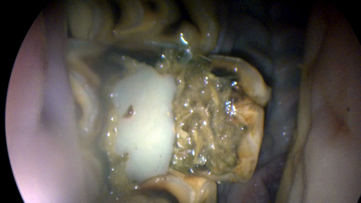
Here is an oroscopic picture of the tooth. As you can see there is a white composite which is the filling material sandwiched between two fragments of tooth. As the fragments have separated, food has travelled up between the two allowing infection to spread to the root of the tooth.
Stuart performed the extraction under standing sedation and a local anaesthetic nerve block which anaesthetises the entire right upper jaw. Initially the gum is elevated from around the tooth. The gap behind and in front of the affected tooth is then spread using appropriately named ‘spreaders’. Forceps are then placed securely on the tooth and gradual left to right movements are made to stretch the periodontal ligament, which secures the tooth within the socket. When loose enough the tooth is then ‘fulcrumed’ (pulled at a right angle) out from the socket. The socket can then be fully examined and cleaned whilst placing a honey soaked swab to help the socket to heal.
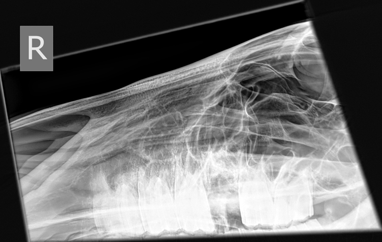
An x-ray taken following tooth extraction shows no remaining root and lots of bony reaction surrounding the socket due to the infection and inflammation that will now subsequently resolve.
Bonita was soon back to eating hay as if nothing had happened. She was discharged home the day after the procedure on antibiotics and pain relief and we are pleased to report that after 4 weeks the socket has completely healed!!!
Horses cope incredibly well with extractions and only usually need 1 week off from ridden work. Bonita’s owners are continuing to keep a close on her for any evidence of further oral discomfort such as quidding and dropping of her feed.