Call Us
Goosnargh:
Lytham Road:
Kirkham:

Sheep – The Big Five diseases

The Big Five Diseases in Sheep

Maedi Visna
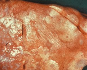
Maedi Visna - sheep diseaseMaedi Visna is a wasting disease causing chronic pneumonia. It is caused by a highly contagious virus. Most sheep are infected as lambs via colostrum or aerosol, however generally do not show signs until they are 4 years old.

The main signs are:

[checklist icon=”fa-arrow-circle-right” iconcolor=”#39aa87″ circle=”yes”]

  • Thin ewes/wasting away
  • Pneumonia
  • Mastitis
  • Arthritis
  • Reduced fertility
  • Smaller weaker lambs which grow poorly due to a lack of milk

[/checklist]

Blood tests are commonly used to diagnose MV. Selecting thin cull ewes to test will help identify the disease if present within the flock.

If you suspect MV in your flock or wish to know your MV status we can do a 12 cull ewe screen.
Please call 01772 861300 to discuss.


Ovine Pulmonary Adenocarcinoma – OPA
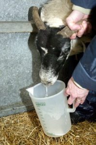
Ovine Pulmonary Adenocarcinoma - OPA - sheep diseaseOvine Pulmonary Adenocarcinoma is caused by a virus leading to a progressive and fatal infectious lung cancer of sheep. The virus is spread from infected sheep by aerosol and nasal discharge. It is often seen in older thin sheep. There is no treatment.

The signs are very similar to chronic pneumonia:

[checklist icon=”fa-chevron-circle-right” iconcolor=”#39aa87″ circle=”yes”]

  • Difficulty breathing
  • Weight loss
  • Excessive watery fluid from the nose
  • Sudden death
  • Secondary infections such as Pasteurella are common.

[/checklist]

Once signs are evident the disease is fatal.

The definitive diagnosis is made by post mortem examination of the lungs.

If there is suspected OPA ultrasound examination of the lungs can be considered.


Johne’s Disease
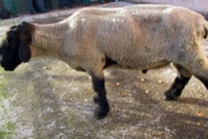
Johne's disease in sheepJohne’s disease is caused by a bacterium which grows very slowly and lives for a long time in the environment. It is mainly spread through faecal contamination of feed and the environment, ewes also pass it to their lambs across the placenta and in colostrum. It is the same agent that causes Johne’s disease in cattle.

Infection generally occurs early in life, sheep often do not show signs for years and act as carriers, these are referred to as ‘sub-clinically infected’.

Affected sheep suffer from:

[checklist icon=”fa-chevron-circle-right” iconcolor=”#39aa87″ circle=”yes”]

  • Severe weight loss
  • Sudden death
  • Anaemia
  • High parasite burdens
  • Affected flocks suffer from poorer production, more thin ewes and increased culling.

[/checklist]

Testing for Johne’s can be tricky at an individual level. There is no single diagnostic test that can detect the disease at all the stages and sub-clinically infected animals may test negative.

Blood screening groups of cull/thin ewes can be effective at identifying Johne’s disease in your flock.


Border Disease
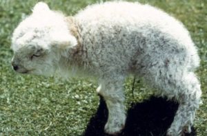
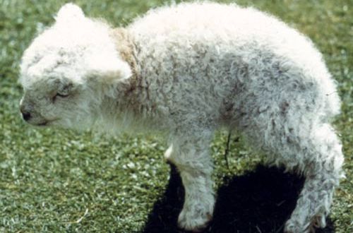
border disease in sheepBorder disease or hairy shaker disease is a virus which causes birth defects, barren ewes and abortion. It is from the same family of viruses as BVD in cattle.

If a ewe is infected during pregnancy the virus passes through the placenta to the lamb, in some cases this causes embryonic loss or abortion. Some lambs survive and are born Persistently infected (PI). PIs shed the virus continuously and can infect other sheep and lambs. This can be a major problem in naïve flocks unknowingly buying in PIs which can then cause around half of lambs born to be affected by the disease.

[checklist icon=”fa-chevron-circle-right” iconcolor=”#39aa87″ circle=”yes”]

  • Infection in non-pregnant adult sheep is generally short lived and mild, often no clinical signs are seen.
  • Hairy shakers are lambs infected with the virus whilst developing in-utero effecting their nervous system which leads to trembling and incoordination. They also have long curly wool.
  • Some lambs are born completely normal but maintain the infection, these are called Persistently Infected animals (PI).

[/checklist]

Hairy shakers are usually a good way to diagnose the presence of the disease. These lambs can also be blood tested to confirm presence of the virus.

Blood testing a group of ewes can identify if the flock has been exposed to the disease and is likely to be present.


Caseous Lymphadenitis – CLA
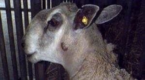
Caseous Lymphadenitis - CLA - sheep diseaseCLA is a chronic bacterial infection of the lymph nodes resulting in abscesses. The bacteria often enters via cuts and is commonly spread through close contact when animals are housed or are trough fed. The disease can also be transmitted indirectly such as on shearing equipment.

[checklist icon=”fa-chevron-circle-right” iconcolor=”#39aa87″ circle=”yes”]

  • Infected animals have lumps often behind the jaw, in the arm pit and in the groin. These lumps are often cold and painless but can burst open and drain pus. Once they have healed and scarred over the abscess can re-occur.
  • Abscesses also form inside the sheep causing ill-thrift and weight loss.

[/checklist]

Lumps and abscesses are a classic sign of the disease and should not be ignored.

Diagnosis is either by sampling the intact abscesses and culturing the pus or by blood test.

If you would like further information, please contact our Farm Team.

Neonatal Lamb Care

Neonatal Lamb CareLambing time is the most crucial part of the year for making your sheep business profitable. Lamb deaths from birth to 3 days old should be less than 7% however many farms range between 10- 25%.

A major factor influencing mortality in neonate (young) lambs is ewe body condition; as this directly relates to lamb body weight, colostrum quality and quantity. It is therefore important to ensure ewes are at BCS 2.5 to 3.0, (so vertebral spinous processes only felt with pressure and transverse processes only felt with hard pressure), and they are on a rising plane of nutrition in the run up to lambing. Energy and protein blood profiles can be taken 2 to 3 weeks before lambing to ensure ewes are receiving the correct nutrition to help reduce cases of twin lamb disease.

Another major cause of neonatal mortality is watery mouth. These lambs typically are lethargic, salivate and have a distended abdomen. It is caused by Ecoli and picked up by ingestion. Treatment involves electrolytes and antibiotics but is often hopeless. Therefore the emphasis should be on prevention which involves keeping pens clean, (especially towards the end of the season), dipping navels with strong iodine and ensuring all lambs get enough good quality colostrum quickly. See below:


Always ensure colostrum is…

QUALITY

Ensure ewes in good BCS, if you’re uncertain as to the quality of colostrum; colostrum quality can be measured using a refractometer: aim for specific gravity >1.05. Only use sheep colostrum replacement (colostrum from a milking sheep farm will usually be better quality than powdered colostrum); cow colostrum can cause a fatal anaemia. Also remember to give your Clostridial and Pasteurella vaccine four to six weeks before lambing to ensure the lambs receive antibodies against these diseases. (Any ewes that haven’t had this vaccine before will need a course of two injections four to six weeks apart).


QUANTITY

200ml/kg within 18 hours of birth, with a maximum of 50ml/kg intake on each occasion.

Remember lambs in adverse weather conditions will require more colostrum or milk as more energy from their feed will be used to keep them warm.


QUICKLY

Within 2 hours of birth.


SQUEAKY CLEAN

Ensure ewe udders are clean and dry and the equipment to mix and administer colostrum is cleaned and disinfected between lambs.


QUANTIFY

We can also look if your lambs have received enough colostrum by blood testing any less than a week old.


Finally, there is continual pressure on antibiotic use. So to ensure we don’t end up with a world full of super bugs we need to be careful how we use antibiotics. Preventative use of antibiotics, such as blanket use of Spectam or Betamox LA for watery mouth needs to be reduced as this will actively select for resistance and there is pressure from Red Tractor Assurance to reduce this practice.

We recommend only using antibiotics….

    • Towards the end of the lambing season, when the sheds have a build up of Ecoli.
    • In triplets, when their ewe may have poorer quality and quantity of colostrum
  • In lambs from ewes in poorer condition, as the quality and quantity of colostrum will be poorer.

Also remember entropion, where the lamb’s eyelid is inverted causing the eye to be held shut, tear staining and damage to the eye. This is a heritable condition so affected lambs should not be kept as replacements, ewes should not be used to breed replacements and consider ram choice if a large percent of flock are affected.

If you have any questions regarding lamb care, please contact the Oakhill Farm Team.

Mastitis in ewes

Mastitis in ewes can be fatal and usually results in the end of her productive life.

The usual presentation is acute gangrenous mastitis (blue or black bag) usually caused by either Staphylococcus Aureus or Mannheimia Haemolytica. S. Aureus is normally present on the teat skin but damage to the teat ends allows it access into the teat canal and causes mastitis.

M. Haemolytica is present in the tonsils of lambs so colonises the udder during suckling. The bacteria produces toxins which prevent blood flow and cause the udder to turn blue/black hence the name. This often occurs around peak lactation (three to six weeks post-lambing) and the initial clinical signs may be as subtle as a ewe looking lame or a lamb bleating because it’s hungry. The disease then rapidly progresses.


The risk factors include:

[checklist icon=”fa-arrow-circle-right” iconcolor=”#39aa87″ circle=”yes”]

  • Under feeding ewes round lambing or ewes in poor condition, as lambs to these ewes will cross suckle or create teat lesions by over suckling.
  • Damaged teat end or Orf on teats
  • Lumps in the udder
  • Lambing inside (the longer the time spent inside the greater the risk of developing mastitis)
  • Dirty, wet pens and dirty hands when handling udder/ underneath of sheep
  • Harsh, cold weather

[/checklist]


Treatment is unlikely to save the udder but may save the ewe’s life:

[checklist icon=”fa-arrow-circle-right” iconcolor=”#39aa87″ circle=”yes”]

  • Remove lambs from the ewe and ideally artificially rear as rearing these lambs on another ewe may spread the mastitis
  • Antibiotic injectable AND non-steroidal anti-inflammatory such as Loxicom
  • A ewe that has had mastitis will be more likely to get fly strike so preventative fly treatment is essential.
  • There is a vaccine against S. Aureus available; VIMCO. This has to be given three and five weeks pre-lambing. Most of the trial work has been done in dairy ewes and it is licenced to reduce to the incidence of subclinical mastitis. Potentially this could also help to reduce the incidence and severity of black bag in your flock however at the moment there is limited evidence to prove this.

[/checklist]

If you have any questions regarding mastitis in your flock, please contact our Farm Vet Team.

Equine Influenza – what’s the current situation?

There have been separate cases of Equine Influenza confirmed in the UK. Horses in Essex, Cheshire and Derbyshire have been affected this month with further reports of outbreaks in Belgium, France and Germany. With all three outbreaks it has been unvaccinated horses that have tested positive.
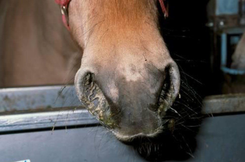
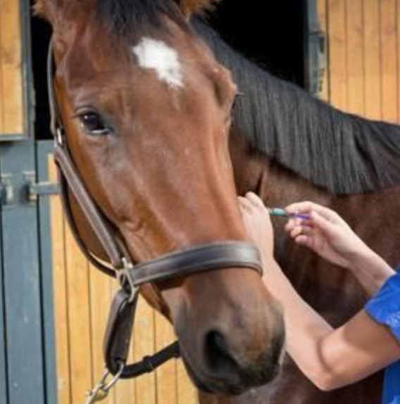
Currently the number of outbreaks is small but they have occurred within a short duration; to put things into perspective a similar number of cases were reported during the whole of 2018.

It is currently unknown which strain of the virus has caused these outbreaks, but research is being conducted to find out. While there are several signs of flu in horses, those affected may only show one or two of them.

What are the signs of infection?

Unvaccinated horses

  • In unvaccinated horses the virus targets the upper respiratory tract, this leads to a very dry, harsh or hacking cough.
  • They usually have a raised temperature which can last around 7-10 days. The fever often makes them dull, off their food and lethargic
  • A clear or white nasal discharge is often seen with enlargement of the submandibular lymph nodes (under their chin/throat area).

Vaccinated horses

  • Horses that have been regularly vaccinated often show no clinical signs, but they may still shed enough virus to infect other horses. This is how the outbreak in Australia in 2007 started.
  • Horses that have been vaccinated but only have partial protection, e.g. because they haven’t been vaccinated frequently enough or because the vaccine type used was not updated, may show varied signs of mild non specific respiratory disease. This can include mild lethargy, nasal discharge and possibly a cough.

What to do if you think there is a possible infected horse?

Owners should seek veterinary advice if they suspect there is flu on the yard and carry out the following precautions:

  • Isolate the possibly infected horses. Infected horses will spread the virus in respiratory droplets, through coughing, and can spread the virus for up to 10 days. They must be isolated until the vet advises they are no longer infected.
  • All horse movements on and off the yard should be stopped.
  • Monitor all horses on the yard for clinical signs and record their rectal temperature daily, it should be less than 38.0˚C. A rise in temperature can be an early sign of an infection.
  • Booster vaccinating all in-contact horses, even if they are not yet due their annual booster, has been shown to provide horses with even more protection against flu.

How to prevent it

Horse Flu is endemic in the UK, which means that most horses will come into regular contact with the virus during their lifetime. The control of horse flu in the UK is based on limiting the signs of infection in horses that have been exposed to the virus, rather than trying to prevent exposure itself. This is primarily achieved through regular vaccination which is given either once or twice a year.
If you have any questions about your horse’s vaccinations, please call our Equine Team on 01772 861300

What lurks beneath – equine lice

As our horses grow a thicker and longer coat over winter to keep themselves insulated it becomes an ideal breeding ground for lice, especially when we then cover this in a warm rug.

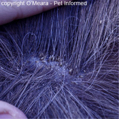
The life cycle of the louse is complete within 4 weeks and eggs hatch 10 days after being laid. Eggs or nits are small (1mm), yellowy-white in colour and glued tightly to the base of hairs.

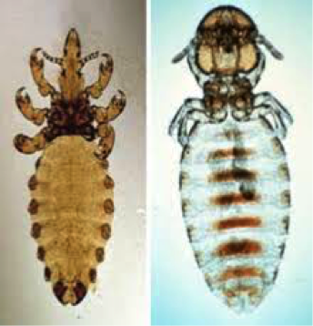
Damalinia

Haematopinus Lice

Haematopinus

All horses can become infected with lice and those kept in large groups indoors, the elderly and young and those with immunosupressive conditions such as malnourishment and PPID (Cushings) are at an increased risk.

There are two types of lice affecting horses, a biting louse called Damalinia equi and a sucking louse called Haematopinus Asini.

Biting lice feed on skin scales and are found where the coat is finer (neck & body) whereas sucking lice feed on blood and are found where the hairs are longer in the mane and tail.

The feeding nature of the lice causes skin irritation with horses becoming extremely itchy. This can lead to patchy hair loss and a scurfy coat. In severe infestations the horse or pony can become anaemic.

Diagnosis is based on evidence and appearance of lice or their eggs.

Removal of the winter coat will help ensure treatment reaches the skin and has the best effect. Lice powders are available over the counter however anecdotally they don’t seem to work. Treatment needs to involve a permethrin or cypermethrin product applied every two weeks for a few treatments to ensure all life stages of the lice are killed. In addition if there is an underlying cause this needs to be identified and treated to prevent reoccurrence.

The lice affecting horses can’t affect humans however sharing rugs/ numnahs/ grooming kits can spread it horse to horse.

If you have any questions regarding lice on your horse, please contact us.

Extended liver fluke season and climate change

Fascioliasis

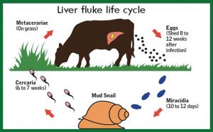
An important stage of the Liver fluke life cycle takes place in mud snails and requires wet and warm conditions (above 7 – 10°C) during the summer months. Over the last couple of years unseasonal weather has seen these conditions well into autumn and winter. This means that sheep and cattle are still being infected later in the season and more frequent testing and treatment may be required.

Fascioliasis can have a serious financial impact on a sheep farm with immediate losses up to 10 per cent caused by acute/subacute disease. Chronic disease could half profits by reducing lamb crop and increasing ewe mortality. Lack of treatment in cattle will lead to reduced growth and yields and discarded milk if treatment is no targeted.

We recommend testing your sheep and cattle for the presence of Liver fluke. Treatment needs to be targeted at your flock/herd and will vary from farm to farm. Please discuss your individual requirements with your vet to develop a treatment plan for the coming months.

liver fluke life cycle

Border Disease

Border disease or hairy shaker disease is a virus which causes birth defects, barren ewes and abortion. It is from the same family of viruses as BVD in cattle.
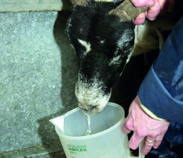
If a ewe is infected during pregnancy the virus passes through the placenta to the lamb, in some cases this causes embryonic loss or abortion. Some lambs survive and are born Persistently infected (PI). PIs shed the virus continuously and can infect other sheep and lambs. This can be a major problem in naïve flocks unknowingly buying in PIs which can then cause around half of lambs born to be affected by the disease.


[checklist icon=”fa-arrow-circle-right” iconcolor=”#39aa87″ circle=”yes”]

  • Infection in non-pregnant adult sheep is generally short lived and mild, often no clinical signs are seen.
  • Hairy shakers are lambs infected with the virus whilst developing in-utero effecting their nervous system which leads to trembling and incoordination. They also have long curly wool.
  • Some lambs are born completely normal but maintain the infection, these are called Persistently Infected animals (PI).

[/checklist]

Hairy shakers are usually a good way to diagnose the presence of the disease. These lambs can also be blood tested to confirm presence of the virus.

Blood testing a group of ewes can identify if the flock has been exposed to the disease and is likely to be present.

To discuss with our Farm Team further, please contact us


Ovine Pulmonary Adenocarcinoma – OPA

Ovine Pulmonary Adenocarcinoma is caused by a virus leading to a progressive and fatal infectious lung cancer of sheep.

The virus is spread from infected sheep by aerosol and nasal discharge. It is often seen in older thin sheep. There is no treatment.

The signs are very similar to chronic pneumonia:

[checklist icon=”fa-arrow-circle-right” iconcolor=”#39aa87″ circle=”yes”]

  • Difficulty breathing
  • Weight loss
  • Excessive watery fluid from the nose
  • Sudden death
  • Secondary infections such as Pasteurella are common.

[/checklist]


Once signs are evident the disease is fatal.

The definitive diagnosis is made by post mortem examination of the lungs.

If there is suspected OPA ultrasound examination of the lungs can be considered.

If you wish to discuss further, please call our Farm Team on 01772 861300

Winter series: Impactions

Colic is a collection of clinical signs shown by a horse indicating abdominal discomfort. We see an increase in colic’s due to impactions during the winter months.

Impactions of the gastro intestinal tract commonly occur at the pelvic flexure. The pelvic flexure is a part of the large colon which turns 180 degrees on itself and naturally narrows. The reduced speed of food material passing through this area mean it is more susceptible than other areas to blockage.

Other than anatomy there are a number of changes that occur, mostly in the winter, which increase the risk of blockage:

[checklist icon=”fa-arrow-circle-right” iconcolor=”#39aa87″ circle=”yes”]

  • We reduce the amount of hours that horses and ponies are outside eating short moist grass and replace it with eating longer, drier roughage such as hay and haylage. Larger amounts of hay and haylage can be eaten alot quicker.
  • Horses and ponies often have less exercise either because they are confined to a stable for longer periods of the day or because we can’t ride them as much due to shorter daylight hours.
  • Horses may reduce their winter intake due to the water being cold or frozen, causing the food material to become dehydrated in the gut.
  • Horses may be turned out on sand paddocks to stretch their legs. If roughage is fed from the floor then they will ingest sand particles which will rest in the gut.
  • Horses and ponies may eat large amounts of straw from their bedding.

[/checklist]

As food material slows down in the pelvic flexure more water is reabsorbed from the gut causing drying of the gut contents, reducing motility of the gut further, leading to more drying and eventual blockage. Usually the horse will have passed reduced amount of, drier than normal, faeces over the preceeding 48-72 hours to eventually passing no droppings at all.

Pain from impaction colics is due to stretch of the gut.  Impaction colics usually are mild in pain but can have periodic episodes of more intense pain. Normal colic signs such as pawing, flank watching and inappetance can be exhibited.

Examination by a vet may reveal a mildly increased heart rate, dry gums and prolonged skin tent. Gut sounds can sometimes be reduced due to slowing of the gut, however they can also be increased as the body’s response to try to shift the blockage. Impactions of the pelvic flexure can usually be identified on rectal examination. They usually are doughy in consistency and sit in the lower left area of the abdomen.

Treatment, as with any colic, involves providing pain relief and a muscle relaxant. In addition the blocked food material needs to be rehydrated. This is most commonly done using a tube passed down the nostril of the horse into the stomach. A mix of water and electrolytes is then administered. Over time this will seep through the food and hopefully relieve the blockage. Food is restricted until poo is seen as it will contribute to the blockage. Tubing may have to occur repeatedly over several days. In severe cases intra venous fluids may need to be used.

So to prevent your horse or pony from becoming impacted this winter try to:

[checklist icon=”fa-check-circle” iconcolor=”#39aa87″ circle=”yes”]

  • Change diet slowly.
  • Avoid straw bedding if you are concerned your horse or pony eats it.
  • Try to keep exercise maintained, a walk around the yard is better than standing still in a stable.
  • Ensure a ready access to fresh water.

[/checklist]

It is worth noting that there are other causes of impactions such as ileo-caecal impactions due to tapeworms and ascarid impaction of the small intestine in youngsters and these will be discussed at a later date.


As always, if you are concerned about colic in your horse, please call us on 01772 861300


Winter Series: Mud Fever

Mud fever, also known as greasy heel syndrome but correctly termed pastern dermatitis, is an infection of the skin usually of the pastern.

The bacteria causing the infection can be from the environment or living on the skin itself and will gain entry due to an injury to the skin surface. It more commonly affects white socked legs, as the skin’s immune function of these areas is reduced. In minor cases it may present as just a few scabs, however, it can spread above the fetlock, occur concurrently with cellulitis, or leg filling, and be openly bleeding.

Although it can have a typical appearance sometimes a vet visit is needed to rule out other conditions and to identify predisposing factors.

The skins integrity is weakened by continuous wetting of the skin and rubbing from mud meaning it is a problem commonly seen in Winter. However, it may not just be wet and muddy conditions that are causing the problem……..

[checklist icon=”fa-arrow-circle-right” iconcolor=”#39aa87″ circle=”yes”]

  • Lymphoedema: this is typical of cob type or draught breeds. Lymph fluid circulates around the body carrying nutrients in lymph vessels. The lymph vessels in these types seem to be less effective in circulating blood in the lower limbs which leads to accumulation of fluid and skin thickening.
  • Leucocytoclastic vasculitis: this is an immune mediated condition whereby blood vessels become damaged. Personal experience shows it occurs more commonly on the outside of white pasterns.
  • Photosensitisation: this is caused by either liver disease or allergic reaction meaning the horse doesn’t deal with chemicals that react with light meaning that when the horse is exposed to UV radiation (sunlight) it becomes sunburnt.
  • Feather mites/ Chorioptic mange: Chorioptes mites causes itchiness of the lower limbs. This can lead to self trauma and wounds allowing skin infection.

[/checklist]

To rule out these conditions it may be necessary to take blood samples or biopsies to determine if one of these are involved.

In order to treat Mud Fever you may need to treat the underlying cause. Sometimes a thorough antibacterial cleaning and removal of scabs is needed, under sedation, so that topical antibacterial, steroidal and protectant creams can be applied directly onto the damaged skin. In some cases when associated with cellulitis or more widespread infection of the leg systemic antibiotics may be needed.

The deliberation is always; do you wash the mud off your horses legs or do you brush it off when dry ?? In our opinion neither is superior. If you are to wash your horses legs they need to be thoroughly dried and if there is already some mud fever present avoid wetting the leg further.

If you are concerned about Mud Fever in your horse, please call us on 01772 861300