Call Us
Goosnargh:
Lytham Road:
Kirkham:

Equine Standing MRI – what to expect

What to expect when your horse has a standing MRI.

Using the right tools early in the lameness process to get a definitive diagnosis will allow you and your vet to devise the right treatment plan, hopefully getting your horse back to full fitness as quickly as possible.

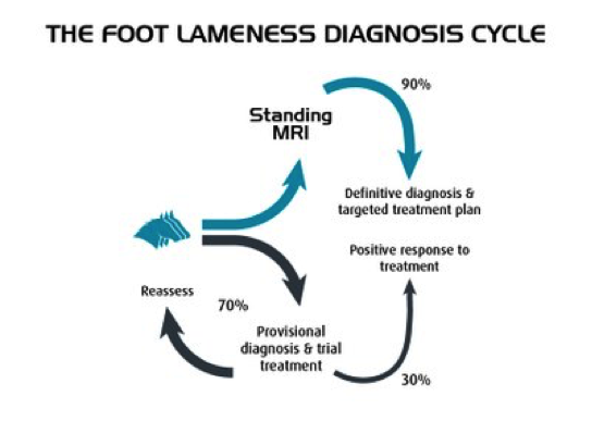
The clear images from MRI allow vets to make an accurate and precise diagnosis in 90% of cases.

If you’ve considered requesting an MRI for your horse but wondered what actually happens, an MRI scan will usually include the following steps:

  1. Initial examination

    On arrival for the scan the horse’s overall health is evaluated for sedation and our clinic vets will briefly examine the horse’s lameness.


  2. Horse shoes

    Metal horse shoes would degrade the quality of the images if left on as the MRI scanner contains a large magnet. Normally just two shoes, on the leg to be scanned and the adjacent leg, are removed.


  3. Sedation

    The standing MRI eliminates the need for anaesthesia, so removes the mortality risk and often allows for day patient scheduling. Top up doses may be applied during the scan, either on a drip or via a catheter in the horse’s jugular vein.


  4. Equine Standing MRIPositioning

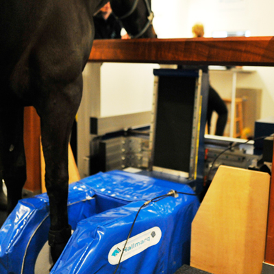
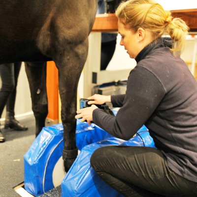
    The horse is walked into the MRI scanner, with the lame leg placed between the poles of the magnet. A radiofrequency coil is fitted around the injury site and the operator makes careful adjustments to ensure the horse and magnet are both in the right place.


  5. The Scan

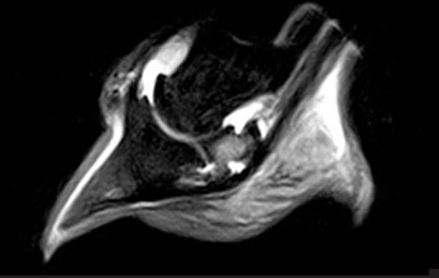
    equine MRIThe scan takes around 2 – 4 hours, producing around 300-500 images at multiple angles of the limb or hoof, highlighting different types of tissue and pathology.


  6. Recovery

    After the scan the horse is given time to recover from the sedation, and in most cases can return home the same day.


  7. Interpretation

    One of our specialists responsible for scanning will carefully review the images to arrive at an opinion about likely pathology or injury. The findings are then communicated to you or your vet.


  8. Treatment

    The findings from the scan will enable an accurate diagnosis to be made. With precise information available the vet can prescribe the best possible treatment for the horse.

Should your horse be suffering with lameness or poor performance issues, please discuss with your usual Oakhill Equine Vet or call the practice on 01772 861300.

If you wish to be referred to us from another veterinary practice, please contact your own veterinary surgeon in the first instance.

Why MRI?

Our equine clinic has the most technologically advanced standing MRI machine available, used for both clients and those referred from other veterinary practices.

Often during a lameness work-up, your vet will use ‘nerve blocks’ to locate where the pain is coming from. This may then be followed with X-ray or ultrasound examinations, but because these imaging tools only show bone or some soft tissue your vet may not be able to see abnormalities, particularly those within the hoof capsule.

Standing equine MRI offers unique insight into equine lameness, identifying the specific cause in over 90% of cases.

With no general anaesthesia required the inherent risks associated with anaesthesia and recovery can be avoided and an early, safe and accurate lameness diagnosis will save you time, money and worry.


We’re often asked….

equine MRIWhat is different about MRI?

MRI images show information about both bone and soft tissue, whereas x-rays only show bone and ultrasound only shows soft tissues. The many, clear images from MRI allow vets to make an accurate and precise diagnosis in 90% of cases.

Foot Lameness Cycle


Oakhill equine MRI referral serviceIs it the same as a human MRI scanner?

The underlying principle is exactly the same but the Hallmarq Standing Equine MRI system has specifically designed for imaging the standing horse, not a human! The scanner operates close to floor level and the horse can immediately step out of an opening in the magnet if it needs to.


Is it safe for my horse?

MRI is widely used in both human and veterinary medicine as it is valued for it’s high image quality of both bone and soft tissue with no ionising radiation. The technology used is unobtrusive and poses no known risk to the horse.

Unlike high-field scanners where the horse has to be anaesthetised, our Hallmarq low-field system uses a smaller magnet that fits around the leg, allowing us to image the horse while standing and under mild sedation.

Hallmarq equine MRI systems have been used for over 60,000 standing sedated horse examinations, during this time there have been no fatalities.


The benefits of having an MRI scan at Oakhill Equine Clinic include:

  • Expert interpretation of images by our ECVS & RCVS recognised surgery specialists, Rosie Owen & Guy Hinnigan and imaging specialist, Meredith Smith.

  • Precise diagnosis

  • Rapid results

  • Specific prognosis

  • Optimum treatment

Using the right tools early in the lameness process to get a definitive diagnosis will allow you and your vet to devise the right treatment plan, therefore getting your horse back to full fitness as quickly as possible.

It is also very useful in establishing an accurate prognosis, to save you time and money worrying about what is likely to happen in the future.

Click here to find out more about what to expect when your horse has a standing MRI.

Should your horse be suffering with lameness or poor performance issues, please discuss with your usual Oakhill Equine Vet or call the practice on 01772 861300.

If you wish to be referred to us from another veterinary practice, please contact your vet in the first instance.