Call Us
Goosnargh:
Lytham Road:
Kirkham:

A SPLINTING NUISANCE – WHEN A SPLINT ISN’T JUST A SPLINT

A SPLINTING NUISANCE – WHEN A SPLINT ISN’T JUST A SPLINT

When is a splint more than a splint? This was the question Paula found herself asking when her horse, Charlie, developed a lump on the inside of his lower leg consistent with a leg splint. However, when the lameness and pain on palpation persisted for several weeks, it became clear that something more serious could be going on.
Horse Charlie's X-ray and MRI scan

Investigations

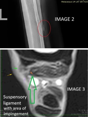
Charlotte, one of our vets, recommended taking X-rays using our ambulatory digital X-ray system at Paula’s yard. The cause of Charlie’s ongoing discomfort was immediately apparent: a section of bone had fractured away from the splint (Image 2, circled). This type of injury is typically caused by trauma, such as a kick from another horse in the field.

Surgical removal of the fractured bone fragment was recommended. Before surgery, an MRI scan was performed to assess any associated soft tissue damage and to help plan the procedure. The MRI revealed that the splint had extended into the suspensory ligament (Image 3), meaning this area would also need surgical treatment.

Surgery

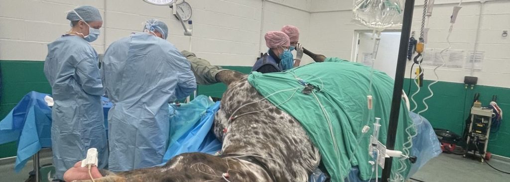
Charlie underwent general anaesthesia, where Guy, one of our specialist surgeons, removed the fractured bone fragment and carefully debrided the portion of the splint that was impinging on the suspensory ligament.

After surgery, Charlie’s leg was bandaged and he began a carefully managed three-month rehabilitation programme. A follow-up MRI confirmed there was no longer any impingement on the suspensory ligament and that the inflammation had completely resolved.
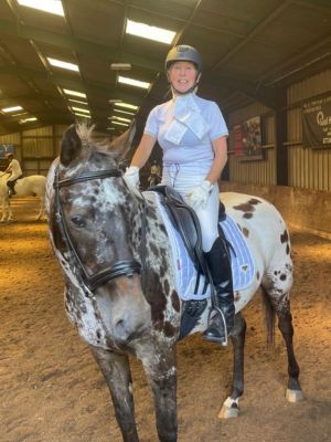
Horse Charlie at a dressage competition

Back to enjoying life!

We’re delighted to say that Charlie has now returned to competition and is enjoying getting out and about again with Paula.

If your horse has a splint that remains painful or is associated with ongoing lameness, don’t assume it’s ‘just a splint’. Our team has access to advanced diagnostic imaging and specialist surgical expertise to investigate these cases thoroughly and help your horse return to full health.

A huge thank you to Paula for allowing us to share Charlie’s story.
SAME NAME, DIFFERENT MANE: WHEN KAI MET KAI!!

CASE STUDY: SAME NAME, DIFFERENT MANE: WHEN KAI MET KAI!!

Kai, a Dartmoor Hill pony, recently had the slightly unusual experience of being treated by his namesake, our vet Kai!

Pony Kai was examined following the sudden onset of severe left forelimb lameness after spending some time turned out in the field.
On closer examination, a small crack was identified along the sole of his foot, which was particularly painful when gentle pressure was applied.

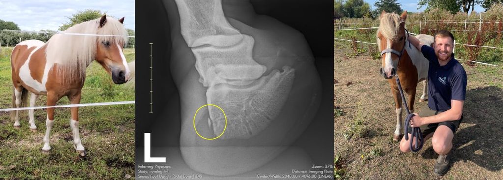
X-rays revealed that Kai had unfortunately sustained a small chip fracture of his pedal bone, with a deep pocket of dirt and small stones trapped beneath the cracked sole.

TREATMENT

Thankfully, the problem could be treated with a standing surgical procedure, which Kai tolerated brilliantly.

The damaged sole was carefully pared away, allowing the trapped stones and debris to be removed.

The affected area was then thoroughly cleaned and flushed, and the contaminated portion of pedal bone carefully debrided.

After a few weeks of box rest, pain relief and antibiotics – along with plenty of bandage changes from his very dedicated owner – we’re delighted to report that Kai has made a fantastic recovery and is now back to his usual self.

And, of course, we couldn’t miss the opportunity for a photo of Kai and Kai, reunited a few days ago under much happier circumstances!

2026 REPRODUCTION SEASON UPDATE

2026 REPRODUCTION SEASON UPDATE

2026 has been another busy and successful year for our Reproduction Team — Rob, Jade and Betty.

Throughout the season, we have performed a variety of frozen and chilled AI procedures, assisted with natural cover cycles, and carried out infertility investigations.

With every mare and client we work with, we aim to gather as much information as possible about the breeding cycle, previous breeding history and semen type selected. This helps us guide you towards the most appropriate approach for your mare.

Every mare is an individual, and we treat each case accordingly. The more we can learn about a mare throughout her cycle, the better we can plan an efficient breeding programme and determine the appropriate level of intervention for her individual needs.

As we head towards the end of summer and the start of weaning, we are still busy with youngstock and foal general health and routine work.

2027 BREEDING SEASON

Preparation for the 2027 breeding season is also already underway. If you are considering breeding next year, or would simply like to discuss your options, please do get in touch with us — we would be very happy to help.

Thank you for your support throughout 2026, and we look forward to seeing you in 2027!

Rob, Jade & Betty

WHAT IS ATYPICAL MYOPATHY?

WHAT IS ATYPICAL MYOPATHY?

Atypical Myopathy (AM) is a serious and potentially life-threatening disease caused by horses ingesting sycamore seeds, seedlings, or leaves that contain a toxin called hypoglycin A. Once ingested, this toxin causes severe muscle damage, affecting the horse’s ability to move, breathe, and maintain normal bodily functions.

HOW MUCH DOES A HORSE NEED TO INGEST TO BE AFFECTED?

Unfortunately, there is no simple answer.

Not all sycamore trees contain the same levels of toxin, and concentrations can vary significantly between trees and from year to year. As a result, there is no known “safe” amount of sycamore material that a horse can consume.

Fortunately, sycamore seeds and seedlings have a bitter taste, and horses generally avoid eating them when sufficient grazing or forage is available. However, young horses can be more inquisitive and may be at greater risk of consuming them.

If you are concerned about sycamore contamination in your paddocks, samples of seeds, seedlings, and leaves can be tested for hypoglycin A. The Royal Veterinary College offers testing services for horse owners wishing to assess the risk on their land.

WHAT ARE THE CLINICAL SIGNS?

Signs of atypical myopathy can develop suddenly and may include:

  • Depression and lethargy
  • Weakness
  • Muscle tremors
  • Stiffness or reluctance to move
  • Rapid of irregular heart rate
  • Dark red or brown urine
  • Difficulty breathing
  • Colic-like symptoms

If you notice any of these signs, particularly during periods when sycamore seeds or seedlings are present, veterinary attention should be sought immediately.

HOW IS ATYPICAL MYOPATHY DIAGNOSED?

Diagnosis is based on a combination of:

  • A history of access to sycamore trees, seeds, seedlings, or leaves
  • Compatible clinical signs
  • Blood tests showing evidence of muscle damage

Additional blood and urine tests can be performed to detect the presence of the toxin and help confirm the diagnosis.

HOW IS IT TREATED?

Unfortunately, there is currently no specific cure for atypical myopathy. Treatment focuses on intensive supportive care and nursing.

This may include:

  • Intravenous fluid therapy
  • Pain relief
  • Nutritional and vitamin supplementation to help limit ongoing muscle damage
  • Careful monitoring and supportive nursing

Early recognition and aggressive treatment are crucial. The chances of survival depend largely on the extent of muscle damage that has already occurred by the time treatment begins.

For severely affected horses, the prognosis remains poor, with survival rates reported between 10% and 25%.

HOW CAN I PREVENT ATYPICAL MYOPATHY?

Prevention focuses on reducing exposure to sycamore seeds, leaves, and seedlings.

Practical steps include:

  • Fencing off areas around sycamore trees
  • Regularly clearing paddocks of seeds and seedlings
  • Providing supplementary hay when grass is sparse
  • Removing sycamore trees from grazing areas where possible
  • Monitoring fields closely during autumn and spring, when the risk is highest

Remember!
Sycamore seeds, often known as “helicopters”, can travel surprisingly long distances. They may be found up to 200 yards from the parent tree, so neighbouring trees can still pose a risk even if there are no sycamores within your paddock.

EARLY ACTION SAVES LIVES

Atypical myopathy is a veterinary emergency. Prompt recognition of the signs and immediate treatment provide the best chance of survival. If you suspect your horse may have ingested sycamore material or is showing any of the signs listed above, contact us without delay.

CELLULITIS VS LYMPHANGITIS- WHAT ARE THE DIFFERENCES?!

Photo Credit: ARND

CELLULITIS VS LYMPHANGITIS- WHAT ARE THE DIFFERENCES?!

A lot of owners are familiar with the terms cellulitis and lymphangitis, but what are the difference in these conditions?

Cellulitis is a bacterial infection of the skin and the subcutaneous tissues. It typically causes a hot, painful swelling in the affected area. Most commonly, it affects the limbs, although it can occur anywhere on the body.

The condition is usually associated with a wound or prolonged exposure to wet and muddy conditions that soften and damage the skin’s natural barrier.
Horses with Chronic Progressive Lymphoedema (CPL) or feather mite infestations can also develop cellulitis.

Clinical signs include:

  • A hot, localised swelling that can be painful to touch
  • Varying degrees of lameness
  • Occasionally, a raised temperature (greater than 38.5°C)

Lymphangitis on the other hand is inflammation of the lymphatic vessels within the limb and is generally a more severe condition than cellulitis.

Affected horses are often extremely lame, have a high temperature, and develop significant swelling throughout the entire limb, extending from the hoof to the elbow or stifle. This dramatic swelling is often described as giving the leg a “tree trunk” appearance.

TREATMENT

Treatment of both cellulitis and lymphangitis typically involves anti-inflammatory medication and antibiotics. Depending on the severity of the condition, your veterinary surgeon may also recommend corticosteroids or intravenous medications.

Prompt treatment is essential. The longer inflammation persists, the greater the risk of damage to the lymphatic vessels, which can become permanent.

In mild cases of cellulitis, horses recover fully and may never experience another episode. However, recurrence is common in more severe cases of cellulitis and lymphangitis due to lasting damage to the lymphatic drainage system.

It is important to remember that not every swollen limb is caused by cellulitis. However, if you are ever concerned about swelling in your horse’s leg, please contact us for advice. Our veterinary team will be happy to discuss your concerns and determine whether an examination is required.

REDUCING THE RISK OF CELLULITIS AND LYMPHANGITIS

While these conditions cannot always be prevented, the following measures can help reduce the risk:

  • Keep your horse moving, as exercise supports circulation and lymphatic drainage.
  • Minimise the amount of time spent standing in deep mud or sand.
  • Thoroughly dry the legs after washing or prolonged exposure to rain.
  • Consider stable bandaging horses that are standing in for extended periods or are prone to limb swelling while stabled provided legs are completely dry before bandaging.
  • Treat for mites if applicable.

Early recognition and intervention are key. If you notice sudden swelling, lameness, heat, or pain in a limb, contact us as soon as possible.

AN EXCITING UPDATE FROM TEAM ANAESTHESIA! 

AN EXCITING UPDATE FROM TEAM ANAESTHESIA!

Environmentally Responsible Anaesthesia

When your horse comes in for surgery, they may need a general anaesthetic to keep them ‘asleep’ and pain-free. Anaesthetic gas (Isoflurane) is essential for safe anaesthesia of our horses but is unfortunately a potent greenhouse gas.  Once the horse breathes out, this gas is released into the air and contributes to global warming by trapping heat in the atmosphere.

Did you know?

Just one hour of anaesthesia can produce 6–8 kg of greenhouse gas. That’s the same carbon footprint as driving 15 to 30 miles in a car!

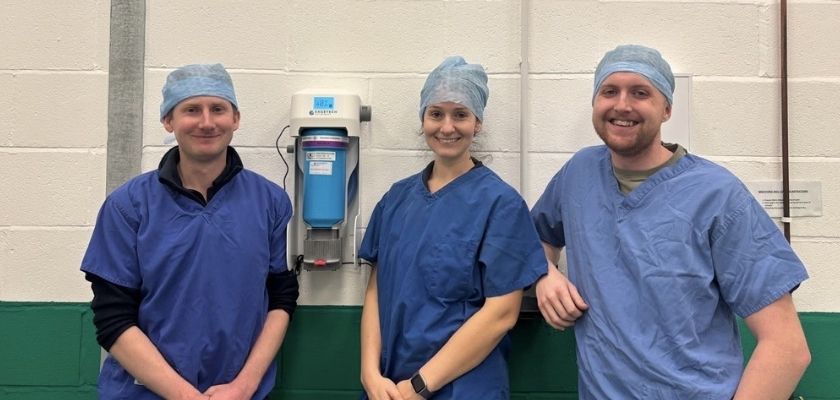
We’ve partnered with SageTech Veterinary, a UK-based company, to install innovative technology that captures and recycles this anaesthetic gas before it escapes into the atmosphere, helping us to lower our environmental impact.  Here is some of our anaesthesia team with our very own ‘VET-Dock’!

This investment is just one of a number of ways that the equine team is choosing to operate in a sustainable and environmentally responsible way when we are delivering clinical service.

If you have any concerns regarding your horse please do not hesitate to contact the team. At least two of our Oakhill Equine Vets are on call 24/7, 365 days/year.

CASE STUDY: DOUG’S STORY

CASE STUDY: DOUG’S STORY

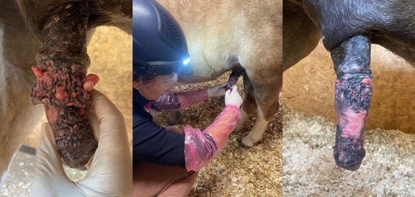
Doug, an 18-year-old palomino pony, was seen by equine vet Jess last August after his owners noticed a build-up of discharge around his sheath that was falling onto his hind legs.

During a closer examination, and following a sheath clean performed under sedation, Jess discovered multiple masses on the body of Doug’s penis. These lesions had the typical appearance of squamous cell carcinoma, a type of cancerous tumour that can affect this area in horses.

The largest masses were removed under local anaesthetic and biopsy samples were submitted for histopathology, which confirmed the suspected diagnosis.

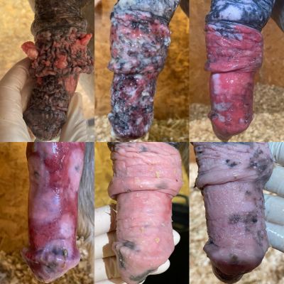
Doug’s owners were keen to explore treatment options that did not involve extensive surgery. A treatment plan was developed using a chemotherapy medication applied directly to the affected areas by Jess over several treatment sessions.

Doug has been an absolute trooper throughout the process. After a few months off he is now back having fun with his lovely family and we are delighted to report that his most recent check-ups have shown an excellent response to treatment – as you can see!

Early investigation of unusual discharge, swellings or lesions can make a big difference to outcomes like Doug’s. If you ever notice anything unusual with your horse, please don’t hesitate to get in touch with our equine vet team.

Thank you to Doug’s owners for allowing us to share his story.

If you have any concerns regarding your horse please do not hesitate to contact the team. At least two of our Oakhill Equine Vets are on call 24/7, 365 days/year.

HIDDEN IN THE HEAD: THE CASE OF A DENTIGEROUS CYST

HIDDEN IN THE HEAD: THE CASE OF A DENTIGEROUS CYST

‘King’, a 2-year-old rescue gelding under the care of World Horse Welfare, was seen by our vet Kai for a persistent discharging tract at the base of his right ear. The issue was clearly bothering him, with frequent headshaking and signs of irritation observed by his carers.

A short course of antimicrobials gave only limited improvement, which raised suspicions that something more unusual was going on. Given the location and lack of response to treatment, a dentigerous cyst was high on the list – a congenital condition involving a tooth that has, quite simply, taken a wrong turn.

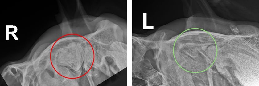
Figure 1 – The red circle highlights abnormal tooth structures in the skull near the ear base on the right side. The green circle highlights normal anatomy on the left side of the head.

King was admitted to our clinic for further investigation, including radiographs and an ultrasound of his head (figure 1). These confirmed our suspicions, revealing a large tooth-like structure lurking deep within his skull – definitely not where you would be expecting to find a tooth! This misplaced tooth leads to a discharge that tracks through the tissues and eventually drains at the base of the ear.

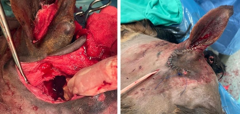
Figure 2 – Photos from during and immediately after surgery.

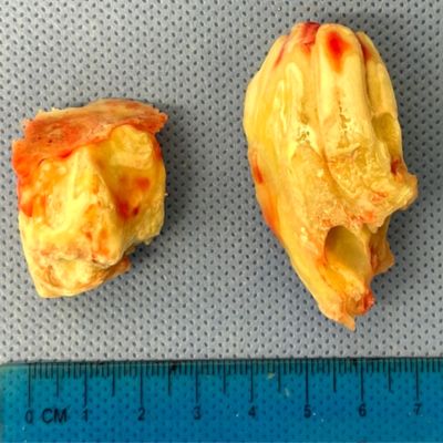
King underwent a lengthy general anaesthetic for surgical removal of the cyst and tooth (figure 2). In a surprising twist, not one but two rogue large teeth were discovered (figure 3). Our diplomat surgeon, Guy, carefully worked over several hours to remove both, all while navigating the many vital structures in the area and avoiding any damage to the nerves responsible for facial movement and ear function.

Figure 3 – The culprits following removal. A 3x2cm tooth and 5x6cm tooth.

We’re very pleased to report that King has since made an excellent recovery. His staples have been removed, he’s much more comfortable, and most importantly, he’s no longer hiding any unexpected dental surprises. We’re delighted to see him doing so well and wish him all the best in finding his much-deserved forever home!

If you have any concerns for your horse, please do not hesitate to contact the team. At least two of our Oakhill Equine Vets are on call 24/7, 365 days/year.

IMPACTION COLIC: SIGNS, TREATMENT AND PREVENTION

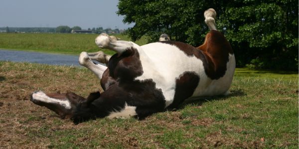
Horse with colic rolling

IMPACTION COLIC: SIGNS, TREATMENT AND PREVENTION

What is colic?

Colic is the term used to describe ‘abdominal pain’ in horses. Fortunately, most colic cases we see are medical colics in particular spasmodic/gas colic. Other forms of medical colic include impaction colic and colon displacements to name but two. Luckily, less than 10% of colic cases we see require surgical intervention.

What are the signs of colic?

  • Restlessness
  • Pawing at the ground
  • Rolling
  • Kicking and/or looking at belly
  • Lying flat out
  • Sweating
  • Stretching/unusual posture
  • Poor appetite
  • Reduced droppings
  • Lip curling
  • Teeth grinding

What should I do if my horse is colicking?

  • Remain calm and keep safe
  • Remove food
  • Walk in hand ideally in a sand paddock provided it is safe to do so
  • Speak to your vet for advice
  • Do not give any pain-relief or other drugs unless instructed to do so by your vet

What is impaction colic?

Impaction colic is a form of colic we see more commonly at this time of year and occurs when gut contents become ‘stuck’ in the colon. Horses generally present with low grade signs of abdominal discomfort but with a history of a reduced appetite and reduced/absent faeces.

How is impaction colic diagnosed?

The diagnosis is based on a combination of historical information, symptoms and veterinary examination. A rectal exam is required to definitively diagnose the impaction.

What is the treatment for impaction colic?

1. Fluids!

  • Water and electrolytes are the best way to help soften and shift impaction colic. These are usually delivered via a nasogastric tube which is passed up your horse’s nose and down into their stomach.
  • Water buffet – ensure your horse has plenty of water to drink
  • Depending on the severity of the impaction repeat tubings is often required at 2–4-hour intervals. Therefore, hospitalisation is required in some cases.

2. Starve!

  • The vet will ask you to remove any hay/haylage/grass. This will stop the impaction getting larger in size.

3. Pain relief

  • Your vet will give your horse injectable pain relief to keep them comfortable whilst we attempt to clear the impaction.

What can I do to prevent impaction colic?

  • Keep up to date with routine dentistry.
  • Ensure your horse follows a worm control plan.
  • Ensure any dietary changes are made gradually over a 10-14-day period.
  • Monitor water intake to ensure your horse is drinking plenty. In winter ensure your horse’s water is not frozen. Consider adding warm water to your horse’s bucket feeds to increase intake. A salt lick should also be provided to stimulate thirst.

If you are concerned your horse is showing signs of colic, please do not hesitate to contact the team. At least two of our Oakhill Equine Vets are on call 24/7, 365 days/year.

ALPHA-2-MACROGLOBULINS – A NEW, EXCITING TREATMENT FOR EQUINE ASTHMA!

Horse with asthma

ALPHA-2-MACROGLOBULINS – NOT JUST FOR JOINTS – A NEW, EXCITING TREATMENT FOR EQUINE ASTHMA!

Clinical signs of equine asthma, formerly known as COPD or RAO, occur when a genetically susceptible horse inhales an allergen to which they are allergic.

This results in airway spasm and an increased production of mucus and inflammatory cells within the airway resulting in a cough, increased respiratory rate and effort and decreased performance to name but a few. The severity of clinical signs vary from mild to severe.

Dust is commonly implicated, but some horses present during the summer months when pollen levels are high.

Diagnosis is frequently based on compatible clinical signs but can be confirmed through airway endoscopy and laboratory analysis of airway fluid samples.

In the past, treatment of asthma relied heavily on bronchodilators and steroids in addition to environmental modifications.

Recently, nebulised alpha-2-macroglobulins has shown promise in the treatment of asthma cases and equine vet Leona has treated a number of our own patients in addition to referral patients using this product.

Alpha-2-macroglobulins are a natural anti-inflammatory molecule which can be harvested from a patient’s own blood, meaning it is a benign and natural product.

In addition to its use in the treatment of joint disease, it can also be nebulised to treat inflammation associated with equine asthma.

Suitable candidates for treatment include those patients where steroids need to be avoided, patients who have not responded to conventional treatment methods or competition horses who require maintenance therapy.

In summary, a really exciting addition to our toolbox for treating equine asthma!

For more information and to see if your horse could benefit from alpha-2-macroglobulins as a treatment for asthma call 01772 861300 to speak to Leona.