Call Us
Goosnargh:
Lytham Road:
Kirkham:

Infundibular Caries

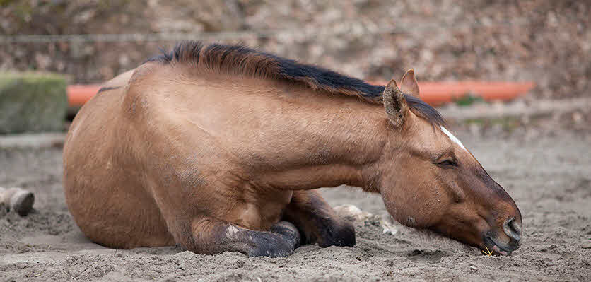
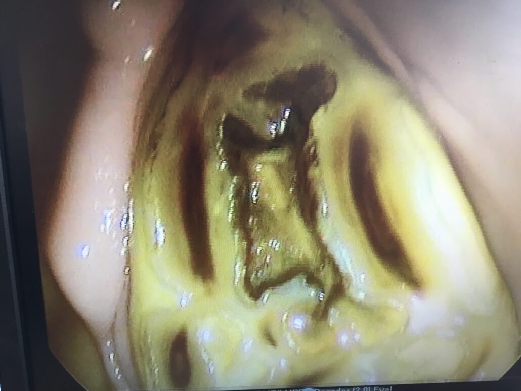
Infundibular Caries is the name given to a condition affecting the upper molars of some horses. Horses have many adaptations in their teeth designed to help with the rough nature of their diet. One of these adaptations is the ‘infundibulum’.

This is simply an extra area of enamel, one of the hardest compounds found in the body, filled with a softer cementum. Having more of this hard enamel is therefore ideal for grinding down rough tree branches in the wild! Occasionally, the area within this extra enamel doesn’t develop as it should, for reasons that are not completely understood.

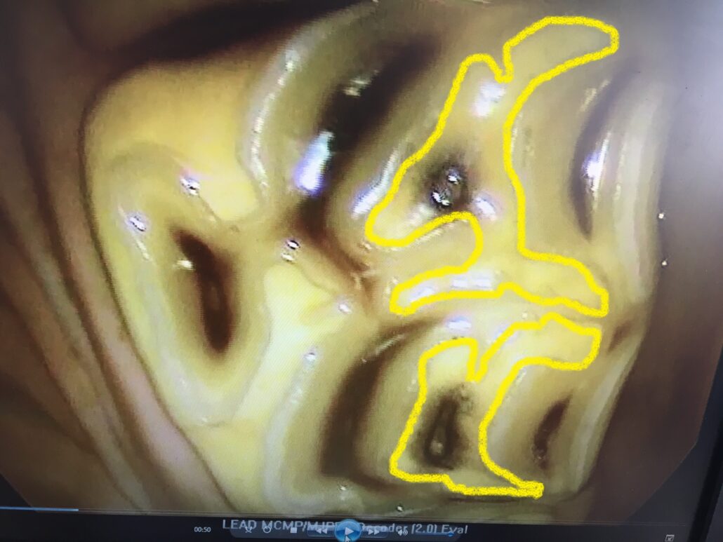
The horse is therefore left with a hole within the tooth which fills with food. Over time this food can cause the tooth to rot resulting in either infection of the tooth or catastrophic fracture. You will be pleased to hear that both of these outcomes are completely avoidable!!

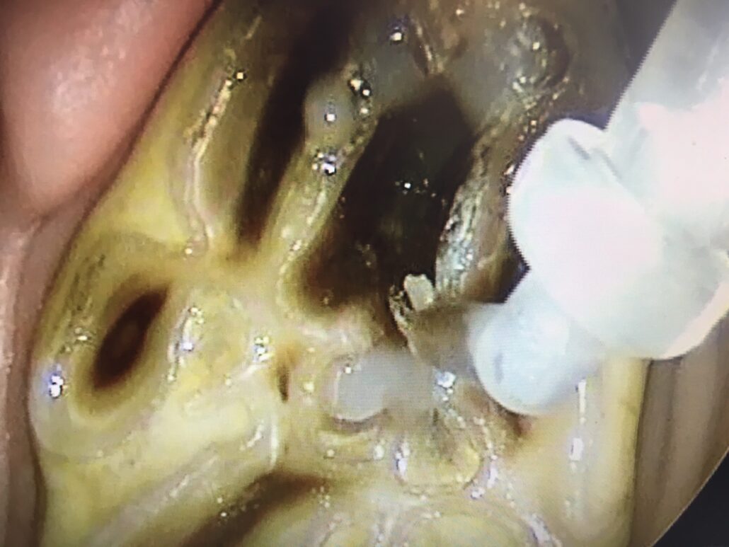
Regular dental examinations using a mirror, or an oral camera, enables us to visualise the surface of the teeth and detect these small but significant defects. Once detected, they can be investigated by probing and/or x-ray to determine the depth of the hole and the requirement for filling.

These holes can easily be filled, much like if you or I went to the dentist! Under standing sedation at our clinic, these holes are thoroughly cleaned out of all the rotten food and filled using a material that binds to the tooth. Once the hole has been filled, food can no longer become trapped and the tooth is no longer at risk of becoming infected or fracturing.

If you have any concerns regarding your horses dental health, please phone our clinic and ask to speak to one of our friendly team who can point you in the right direction! 

Winter Conditions

Colic

  • Colic is a collection of clinical signs shown by a horse that indicate abdominal discomfort.
  • Changes in management, especially if not made gradually, can precipitate episodes of colic.
  • At this time of year, horses undergo a significant management change, coming in from pasture to spend prolonged periods of time in their stable.
  • Therefore, we frequently see an increased incidence of colic cases.
  • Fortunately, the vast majority of cases can be managed medically and although arguably, spasmodic colic remains the most prevalent form of colic that we see, impaction colic occurs at an increased frequency.

Impactions

  • Impactions generally occur following the accumulation of food material, which subsequently dehydrates (dries out), in a horse’s colon.
  • The colon’s pelvic flexure is the most common site for this to occur. The pelvic flexure is a part of the large colon which turns 180 degrees on itself and naturally narrows. The reduced speed of food material passing through this area means it is more susceptible than other areas to blockage.
  • Other risk factors for impaction development include reduced grazing and increased hay/haylage in the diet, reduced exercise due to increased time spend confined in the stable, reduced water intake during cold/frosty periods and straw ingestion (if bedded on straw) to name but a few.
  • Clinical signs of impaction colic include passing reduced amounts of, drier than normal, faeces over the preceding 48-72 hours to eventually passing no droppings at all, reduced appetite and signs of colic which vary in severity but can be relatively mild.
  • Veterinary examination may reveal a normal/mildly increased heart rate, dry gums and prolonged skin tent. Gut sounds are generally reduced. Impactions of the pelvic flexure are confirmed on rectal examination. They are usually ‘doughy’ in consistency and sit in the left, lower abdomen.
  • Treatment, similar to other forms of colic, includes the administration of pain relief and smooth muscle relaxants. In addition, the blocked food material requires rehydration. The most effective way to achieve this is by regularly administering an electrolyte solution via stomach tube. The administered fluid stimulates colon contraction and the fluid passes through the gastrointestinal tract to rehydrate the impaction stimulating it’s passage through the remainder of the gastrointestinal tract prior to evacuation from the body. For the majority of patients, monitoring and treatment of the impaction is carried out most effectively at our equine clinic. Depending on severity, impactions can take varying periods of time to resolve from 24-48 hours to 5 days. Food is withheld until the impaction has cleared and is gradually reintroduced starting with walks to grass and ‘sloppy’ bucket feeds.

Tips for preventing impaction colic (and colic in general)!

  • Make dietary changes slowly over a 2-week period of time.
  • Avoid straw bedding if you are concerned your horse or pony eats it.
  • Try to maintain exercise. A walk around the yard is better than standing still in a stable.
  • Ensure a ready access to fresh water- break ice regularly when temperatures drop- a tennis ball/apple placed in water may help reduce freezing. 
  • Ensure your horse is drinking. Water intake can be increased by adding water to your horse’s feeds. Providing a salt lick will also stimulate thirst.

Mud Fever

  • Mud fever is an infection of the skin usually affecting the pastern.
  • The bacteria implicated (Dermatophilus congolensis), gains entry to the skin most commonly, following prolonged periods of wetting/standing in mud.  
  • The resulting dermatitis is characterised by hair loss and crusting. Hair comes away in tufts bound by a scab/crust (known as paint-brush lesions).
  • Minor cases may present as just a few scabs; however, cellulitis (subcutaneous skin infection) can ensue.
  • Treatment is based on removing your horse from the wet/muddy conditions. Mild cases can be managed topically using a combination of anti-bacterial and steroid based creams. The affected area may also be clipped and cleaned using a warm dilute chlorhexidine solution. Different vets vary in their recommendations as to frequency of cleaning so please follow your vet’s recommendations. In moderate to severe cases, sedation may be required to examine, clip and clean the area in the first instance. In cases with secondary cellulitis, antibiotics and anti-inflammatories are also required.

Rain scald

  • Rain scald is similar to mud fever, in that it is caused by the same bacterium following prolonged skin wetting, but affects the horse’s topline.
  • Treatment mirrors that of mud fever.

Microchipping Rules Oct 2020

Are you organised following the new microchipping rules that came in on 1st October 2020? Now all horses, irrespective of age should be microchipped and registered with the government central equine database. You can check if your horse is registered at www.equineregister.co.uk and follow our flowchart to check that you have satisfied the new guidelines.

If your horse needs a microchip, our equine vets can effortlessly insert one at our clinic or your yard for the price of £25 (plus VAT).

Microchips, does your horse need one?

Horse microchips or transponders are the size of a grain of rice, and are usually placed on the middle third of the crest of the neck. They contain a small computer, which transmits a 15-digit code to a reader when scanned. This code is unique, and can be used to identify an animal throughout their life.

Since July 2009, all horses must have a microchip when they are issued a new passport, however; for horses that had a passport issued before 2009, they may have a passport without a microchip. As of October 2020, by law, all horses must have a microchip, meaning that horses over 11 years of age which do not have a microchip, will need one inserting.

The change in rules has coincided with the creation of the central equine database, which is accessible at www.equineregister.co.uk. All UK passport-issuing organisations (PIOs) have provided this central database with the microchip numbers of horses that are registered with them. This is an important step aimed to centralise all of the collected microchip data. In the event of a lost or stolen horse (every owner’s worst nightmare, I think we will all agree), if a vet scanned a microchip, they would have to ring around each PIO to find which one held the owners details. As there are over 50 separate PIOs, this isn’t an easy task! Now a horse can be scanned, checked on the equine register, bringing up the details of which passport organisation has the owners details, therefore reuniting the animal with the owner without delay.

How do I comply?

First check if your horses has a microchip, this can be found in the passport, and is usually labelled transponder code, or on the markings page underneath a barcode. If you are unsure, one of our vets can scan your horse for a microchip.

If your horse has a microchip, then check it is registered at www.equineregister.co.uk . If it is, you are compliant with the law. If not, you may need to contact your passport agency to update them with the number.

If your horse doesn’t have a microchip, our equine vets can easily implant one by a quick injection. Only a vet is legally allowed to microchip a horse. Once inserted you will need to inform your passport agency of the microchip number (some require a form completing by the vet, some ask for the passport to be sent back for updating).

Private Microchip Databases

One final comment is about private databases such as PETtrac, Petlog or Identibase, which also record pet microchips. These link the microchip number to an owners phone number, and often have 24/7 phonelines, useful if your horse is lost out of hours! It is not a legal requirement however to be registered with these private databases. When we microchip you horse, we are able to register the chip number to the phone number on your account with PETtrac.

Simba the German Shepherd

Simba is a 5 year old German Shepherd dog who has been visiting Oakhill since he was a puppy. In June 2018, he was diagnosed with hip dysplasia. This is a common condition affecting German Shepherds, Labrador Retrievers and Border Collies amongst other breeds. 

The hip socket is more shallow than usual, and the top of the femur (thigh bone) becomes abnormally shaped and doesn’t sit well within the socket. The resulting joint instability can eventually lead to painful arthritis. It was recommended that Simba start joint supplements and hydrotherapy to build muscle and maintain mobility and strength in his hind legs to reduce the likelihood of arthritis in the future.

In May 2020, Simba became uncomfortable on his left hindlimb. He was diagnosed with disease of the stifle (knee) joint. As Simba was overweight, the pressure put through his hindlimbs was significant. It was advised that he lose weight to reduce the burden on his joints. 

Simba’s owner kept a food diary for one week, so that the nurse could calculate how many calories he was eating compared to how many his body required. The nurse then advised a low-fat food and exactly what size portions he required to lose weight healthily. Simba’s owner started the new diet, being careful to reduce any further treats given. 

Simba was regularly weighed, and has lost 20% of his bodyweight. As you can see, he looks fantastic! His owner reports that he’s now full of life, dragging her to be taken for walks and is building up muscle really well at his hydrotherapy sessions.

We’re all so proud of Simba’s progress. If your pet is overweight, our nurses would be happy to talk over the phone about what can be done to help.

Losing weight reduces the burden on joints and helps pets to live longer.

Autumn: Seasonal Advice for Your Pets

As the temperature drops and it gets dark earlier during the autumn months, it’s important to keep our pets safe.

Below are some seasonal hazards to pets for you to be aware of…


1. Conkers

Conkers

While serious cases of poisoning are rare, ingestion can cause marked gastro-intestinal signs such as drooling, retching, vomiting, diarrhoea and abdominal pain. The conker’s case and conkers themselves can also cause intestinal blockages. Dogs usually vomit any ingested conkers quickly so treatment to control vomiting may be needed.


2. Rat poisons

Most, but not all, rodenticides in the UK contain anti-coagulant compounds that interfere with a rat’s ability to clot its own blood. One-off exposure to products bought in garden centres often does not cause problems. However, repeated exposure to products or exposure to professional rodent baits can cause disruption to a pets’s blood clotting ability and result in massive haemorrhage (bleeding). The effects may be delayed for several days and blood-clotting (coagulation) tests are often needed to determine if a pet is at risk of developing problems. Treatment involves giving an antidote and in severe cases transfusions of plasma or whole blood.


3. Luminous necklaces and glow sticks

The chemical mixture within luminous jewellery and glow sticks (often seen at Halloween & Bonfire night) is very irritating to the gums — commonly causing dribbling, frothing and foaming at the mouth, vomiting and stomach pain. Thankfully, although the signs can look dramatic, ingestion is unlikely to cause significant problems, but your pet may require supportive treatment.


4. Oaks and acorns

Exposure to acorns in dogs is common in the autumn and winter.  The toxic ingredient is thought to be tannic acid, which can cause damage to the liver and kidneys. Signs include vomiting, diarrhoea, abdominal pain and lethargy.  Ingested acorns can also cause an intestinal blockage.


5. Fallen leaves

Piles of leaves can develop bacteria and mould.  If your dog ingests these it can lead to gastrointestinal upsets.


6. Antifreeze

cat anti freeze vets preston

The worst of all the chemical spills is antifreeze (containing ethylene glycol), which can leak from a car’s radiator. Ethylene glycol ingestion is very dangerous. It is sweet tasting and very palatable and even a small quantity can cause serious kidney damage and be fatal. The first signs of intoxication can be that your pet appears ‘drunk’.  If you know your pet has ingested ethylene glycol or you have any concerns, contact your vet without delay.  The prognosis becomes less good the longer the delay between ingestion of the antifreeze and initiation of treatment.


7. Chocolate

chocolate

Chocolate sales rise around Halloween but remember it contains a stimulant called theobromine that’s poisonous to dogs.  The amount of theobromine differs depending on the type of chocolate — dark chocolate has the most in it.  Theobromine mainly affects the heart, central nervous system and kidneys.  Signs will occur from 4-24 hours following ingestion and you may see vomiting, diarrhoea, restlessness, hyperactivity, rapid breathing, muscle tension, incoordination, increased heart rate and seizures.


8. Keep safe in the dark

Always wear reflective clothing to make yourself visible when walking near roads in the dark. And don’t forget your pets! Fit your dog and cat with a reflective or LED collar – but make sure only a quick release collar is used for a cat.


9. Fleas

Turning up your heating in your home as the temperature drops in autumn can provide the perfect temperature for fleas. It’s important to treat your pet for fleas all year round. Please contact us if you wish to discuss the various flea treatments available.


10. Fireworks

The firework season seems to be getting longer each year and can be a stressful time for all pets. Read our article on how you can make fireworks less frightening for your pet: https://www.oakhill-vets.com/fireworks-pets/


If you are concerned that your pet may have been affected by any of the above hazards, please call your vet.

Conker poisoning in dogs

Conkers (seeds/nuts of the horse chestnut tree), may be synonymous with autumn but did you know that they can pose a serious risk to your dog if ingested.

Conkers contain a poison called aesculin which is toxic to dogs. A dog would normally need to ingest several conkers to suffer severe poisoning.

Clinical signs are usually seen between one and six hours after ingestion, although they can be delayed for up to two days.

Symptoms of conker poisoning include:

[checklist icon=”fa-paw” iconcolor=”#39aa87″ circle=”yes”]

  • Vomiting, which may contain blood
  • Diarrhoea
  • Drooling
  • Abdominal pain
  • Increased thirst and reduced appetite.
  • Signs of restlessness, wobbliness and muscle tremors may also be seen.

[/checklist]

Poisoning is not the only risk – these large nuts could cause a blockage in your dog’s intestines.

Although fatalities in cases where dogs have consumed conkers are thankfully rare, it is still very serious, so if you suspect your dog has ingested conkers, please contact your vet immediately.
https://www.high-endrolex.com/18
If necessary, the vet will give the dog medication to make them vomit and they may also perform gastric lavage (wash out the stomach).  This is designed to ensure as much of the poison is removed from the dog’s system as possible.

Although dogs love to forage when they’re out exploring, do keep a watchful eye on them when they’re around conkers.

EHC Online (EHCO) – What’s New?

Export Health Certificates Online is a new web-based service for the application and issuing of EHCs from GB to third countries. We have been dealing with this new system for a number of months on a limited number of certificates but as of 21/08/2020 the service has been extended to a greater number of EHCs. Online application for EHCs that are available through EHCO is now mandatory and the number of EHCs available via EHCO is likely to increase significantly in the next 12 months.

The list of EHCs that need to be applied for through EHCO as of 03/08/2020 can be found here: https://www.oakhill-vets.com/wp-content/uploads/2020/08/EHCO.pdf

To summarise the process:

  1. The exporting company needs to register as an ‘exporter’ via the Government Gateway portal.
  2.  The exporter then makes an online application for the relevant EHC – unlike previously there is no PDF to email to APHA and no EXA form to complete although you will have to fill in a series of questions similar to those found on the old EXA forms.
  3. Select Oakhill as the ‘certifier’
  4. As the exporter you will be able to view all your applications on the online dashboard (a similar dashboard is available to us to view all the EHCs that have been assigned to us). In addition, each application should have a status assigned depending on whether it is being processed by APHA (Processing), sent to Oakhill (Dispatched) or signed off by us following completion (Completed).

The actual inspection and certification process that then involves us is very similar to before. A signed and stamped original EHC must still accompany the consignment and we need to keep a ‘certified copy’ of all EHCs at the practice.

An additional change worth noting is that ‘Cancel & Replace’ certificates will now need to be requested by the certifier rather than the organisation that applied for the original EHC.

Further information is available through the APHA website: http://apha.defra.gov.uk/official-vets/Guidance/exports/ehc-online.htm

Magnetic Resonance Imaging (MRI)

Magnetic Resonance Imaging (MRI) is an advanced imaging tool that is widely used in the veterinary and human healthcare fields. MRI has been developed for use on our equine patients and has been found to be invaluable in helping us to attain specific, accurate diagnoses for our patients. Did you know? Using MRI early on in a lameness case can actually reduce your overall spend by achieving a prompt diagnosis and treatment plan without trying numerous other approaches which are unlikely to help in the absence of a specific diagnosis. 

At Oakhill, we have a standing low-field MRI scanner which means that horses are sedated and stood up for the whole procedure, no general anaesthetic is required, YIPPEEE! The use of the standing MRI scanner is now considered the optimum tool for diagnosing orthopaedic conditions in the horse. The MRI machine is a low field scanner and the strength of the magnet is 0.27Tesla, to put this in comparison to a high field scanner used in hospitals which is 1.5Tesla in strength. This is reflected in the size of the magnet, and the ‘U’ shaped standing magnet is much smaller than those in human hospitals. It’s U-shaped and not a cylindrical shape so that the horse can escape easily from the magnet without harm if they suddenly become anxious during the procedure. 

Usually a horse has been through a lameness investigation including nerve blocks prior to MRI. This may then be followed by radiography (X-rays) and an ultrasound scan, depending on the area to be imaged. If there is still no definitive diagnosis after these imaging modalities, then MRI is often advised. Nerve blocks are essential in determining exactly which site we need to scan. 

So, what will happen if your horse requires and MRI scan? Horses are often admitted as a day case. They will have their shoes removed and a catheter placed to allow small increments of sedation at short intervals to be given easily during the scan. A scan of both front feet typically takes 2-4 hours, but this hugely depends on how well the horse stands within the magnet. 

Our MRI machine is positioned within a specially insulated cabin (called a module!). These modules are made specifically for the MRI machine, to ensure control of temperature and signal interference. Occasionally a French radio wave of a certain frequency has been known to disrupt our scanning! The magnet works within a finely monitored temperature-controlled space and is quite sensitive to change. 

The foot is the most common site that we scan with the MRI machine due to the tough hoof capsule making imaging of the soft tissues difficult. We can assess the bones through radiography, however, MRI has a further advantage of being able to assess for fluid (inflammation) within a bone which no other imaging modality can visualise. Since MRI has been being used, ‘Navicular disease’ has become an outdated term, and we now know there are many different soft tissue and bone pathologies encompassing navicular syndrome, which can be individually assessed and a more accurate guidance on prognosis of the horse’s career given through the use of MRI. 

The magnet can also scan other aspects of the lower limb, up to and including the lower carpus (knee) and hock. This can be particularly useful in areas that can be difficult to fully assess with other imaging modalities such as the proximal suspensory ligament. MRI has revolutionised our ability to achieve a fast, accurate diagnosis and has dramatically enhanced our treatment capabilities of the lame horse. 

Liver Case Study

The liver has multiple responsibilities including nutrient and toxin absorption. It has a large ability to cope with insults however, clinical signs will become apparent when 60% of the liver is damaged. 

Signs can include jaundice (a yellowing of the gums or white of eyes), weight loss and in severe cases neurological signs such as head pressing and blindness. 

This pony was presented to vet Stuart after developing excessive sunburn. Unfortunately, a large proportion of his body was white haired so this sun burn covered his face, muzzle, his legs and around his eyes as seen in the pictures below. Blood evaluation showed an increase in liver enzymes (indicating liver damage) as well as bile acids, indicating impaired liver function.

Hairloss and reddening of the skin on face and legs

A liver biopsy was performed with the pony under sedation and local anaesthetic. This ultrasound scan shows the appearance of the liver.

The results of the biopsy showed the pony to have severe fibrosis (scarring) of the liver, due to ragwort toxicity. However, the changes observed were deemed manageable. 

The pony was placed on steroids, to reduce liver inflammation, and antibiotics for the secondary skin infections. He has had regular antibacterial baths and creams applied to sooth his skin whilst avoiding sun contact. 

He has shown a great improvement whilst on medication and as you can see from the pictures his skin is less inflamed and has started growing hair again as seen in this picture.

His bloods will be monitored going forward. We will keep you updated on his progress.  Liver biopsies are simple to perform and gives a great deal more information on how to treat the liver disease affecting your horse or pony.

If you are concerned about anything discussed then please call us and speak to a vet on 01772 861 300 

Pinworm

Pinworm (Oxyuris equi) infection is manifest by affected horses itching their peri-anal and tailhead regions. Adult worms reside in the large intestine with the female worm travelling down the small colon to lay eggs on the skin surrounding the anus (often indicated by a yellow discharge as seen in photo one below). This usually causes intense irritation manifest by rubbing. Occasionally, mature worms are visible protruding from the anal sphincter which we can also see in this photo. 

Diagnosis is confirmed through microscopic examination of a sellotape impression taken from the peri-anal skin or yellow discharge (if present) which will reveal the pinworm eggs (seen in photo two below). 

Most commonly used wormers are effective for pinworm (ivermectin, avermectin or pyrantel based wormers) and should be administered to clinical cases. The peri-anal region and underside of the dock should also be washed using dilute hibiscrub to remove the eggs. We generally recommend cleaning for three to five consecutive days. The stable should also be thoroughly cleaned and disinfected to endeavour to remove environmental eggs.

Pinworm infection should always be considered in cases of tailhead itching but it is also important to consider other potential causes including sweet itch, other allergies, mites and lice. It is also important to remember that pinworm infection is generally not indicated on faecal worm egg counts as the eggs are deposited on the skin surrounding the anus. Therefore, horses with negative faecal worm egg counts may still succumb to pinworm infection.

As always, should you have any questions, please do not hesitate to contact the equine team.