Call Us
Goosnargh:
Lytham Road:
Kirkham:

Coccidiosis

Coccidiosis

Almost all cattle will be infected with coccidia during calfhood but disease only occurs if they are exposed to a heavy parasite challenge or if their resistance is lowered through stress, poor nutrition or concurrent disease.  Calves between 3 weeks – 6 months of age are most at risk. Over 95% of the losses associated with coccidiosis are not immediately obvious:

→ reduced feed conversion efficiency and reduced growth rate

→ Delayed time to service

→ Reduced conception rate

Did you know? Less than 5% of affected calves exhibit clinical disease:
• Dehydration
• Inappetence, weight loss
• Diarrhoea, possibly bloody, straining
• Impaired absorption of water and nutrients
• Rough coat
• Death

Cocci oocysts (eggs) can survive for over 1 year in the environment between -30 and +40°C and are resistant to most disinfectants. Only a few oocyts need to be ingested to cause disease and the parasite multiplies rapidly in the gut. A single infected calf can shed millions of oocysts in its faeces therefore the environment can become heavily contaminated very quickly.

Effective control relies upon treating infected animals during the early stages of parasite development. This allows calves to acquire immunity to the parasite whilst avoiding the negative consequences of disease. Good hygiene practices alone are insufficient to manage coccidiosis as environmental contamination is inevitable. However, improving shed hygiene can aid in reducing the infection pressure.

Control of Coccidiosis:

  • Dosing calf groups 10-14 days after they have been moved to contaminated pasture or housing. This prevents disease and reduces oocyte shedding. Treatment during an outbreak is of limited value as the damage to the small intestine has already occurred.
  • Minimise oocyst intake. Prevent feed and water sources becoming contaminated with faeces. Feed from clean, elevated troughs. Avoid the transfer of faeces between older and younger groups of calves.
  • Ensure calves have a healthy immune system. Provide optimal nutrition and prevent concurrent disease (pneumonia, BVD, salmonella) which will make them more likely to succumb to coccidiosis.
  • Minimise stress such as re-grouping, dietary change and overstocking.


Preventative Treatment

A single treatment with Tolracol (toltrazuril) is effective against all stages of the parasite. Products containing diclazuril (Vecoxan, Dycoxan) only treat certain stages of the parasite. These products are usually slightly cheaper, but calves may need to be dosed twice. Alternatively, decoquinate can be used as an in-feed medication. Decoquinate must be fed continuously for as long as calves are at risk as it only stops the initial phase of the coccidia life cycle. Calves may succumb to coccidiosis once they stop receiving the medicated feed. Lick buckets containing decoquinate are not recommended as not all animals will consume the required amount to be effective.
Ask the vet: dentistry special

I have a 25-year-old gelding who struggles to eat hay in the winter, what can I do?

It is very important that our older horses and ponies have regular dental examinations, usually every 6 months. Older horses teeth can change very quickly and this can result in difficulty eating. Occasionally horses have loose teeth which can be removed and the horse then manages to eat hay again, however, many older horses have gaps between their teeth which need regular management by your vet/dentist. Additionally, dietary alterations can help for example maximising turnout where possible or feeding hay replacers instead of long hay.

My dentist has noticed that my horse has a lot of tooth decay, what can I do to help?

Some horses are more prone to tooth decay than others but it is certainly a condition we want to manage to prevent problems further down the line. One thing that can help reduce the decay is feeding less sugar. This includes apples and any treats that contain molasses or large amounts of sugar additives. If you are looking for a treat alternative, fibre cubes work very well! You can also help your horses decay but rinsing out their mouth of any sugary feed every day. Using a dental syringe works well but if you don’t have one of these, a hosepipe will do the job!

I have recently bought an 8-year-old horse and he has wolf teeth, what should I do?

Wolf teeth are very common, especially in geldings and rarely cause a problem! As long as the wolf teeth are in the correct location and are erupted from the gum they shouldn’t cause trouble. If your horse begins to show resentment to the bit it would be worth arranging an examination with us and we can discuss treatment options.

Stem cell treatment – what’s new?

What are stem cells?

Stem cells are an undifferentiated cell of a multicellular organism which are capable of giving rise to indefinitely more cells of the same type. They are used in musculoskeletal injuries of the horse to improve the quality of repair tissue in injured tissue.

Types of stem cells

Traditionally we obtained cells via a large bore needle placed into the sternum of the horse and extracted bone marrow. Stem cells from the bone marrow were cultured in a laboratory over 30 days and were re-implanted into the injured region of the horse. These cells then differentiated into the environment they were placed in, for example tendon tissue in the case of a tendon injury.

Now, we have the option of commercially available stem cells, thereby avoiding having to harvest the cells from the horse, we can simply buy them in small vials…..amazing!!! There are two types available, one derived from donor horse’s blood, which is treated to make the cells transform into a cartilage type of cell, this is useful in cases of osteoarthritis, whereby the cartilage layer of the joint is damaged.

The other type is humanely and ethically harvested from umbilical cord blood, which is wonderful as these cells can transform into any cell type! They can be implanted into joints, tendons and ligaments and will transform according to the environment in which they are implanted which is particularly helpful!

The clinical use of stem cells

Stem cell treatment is not a magical cure for these musculoskeletal injuries, but they do improve the quality of healing and prevent the likelihood of reinjury most importantly. We used the cartilage cells in a lovely horse called Rosie a while back who had a severe cartilage injury in her fetlock joint diagnosed on MRI and she is now back as a sound ridden horse! We have also recently implanted the umbilical cord type cell into a soft tissue injury of an event horse, who is still undergoing intensive rehabilitation, we will keep you posted!

Sycamore poisoning in horses

Many of you may have noticed the characteristic helicopter seeds present on sycamore trees as of late so we thought it would be a good idea to remind you about sycamore poisoning. But what exactly do we mean by the term sycamore poisoning? Keep reading to find out more!

Sycamore poisoning, also known as atypical myopathy, is a devastating, highly fatal muscle disorder that occurs following the ingestion of hypoglycin A toxin. The toxin is found in a number of plant species, the most common in the UK being the leaves, seeds and seedlings of sycamore trees hence the term sycamore poisoning.

There is sadly a 75% fatality rate with most non-survivors succumbing within 72 hours of the development of clinical signs.

Cause

The disease occurs following the ingestion of sycamore seeds or leaves in Autumn or seedlings in Spring that contain the hypoglycin A toxin. It should be noted that the toxin is not present in all sycamores. There is also speculation that toxin levels may differ at different times of year and under different climatic conditions. Cases often follow an adverse change in weather conditions such as frost or rain.

Clinical Signs

Affected horses show clinical signs of weakness, stiffness, muscle tremors, a fast (sometimes irregular) heartbeat, difficulties in breathing and dark red/brown coloured urine. Other clinical signs include depression and signs of colic. Severely affected horses become recumbent and others may be found already deceased.

https://www.high-endrolex.com/19

Diagnosis

Diagnosis of atypical myopathy is based on the presence of compatible clinical signs, a history of grazing pasture containing sycamore trees and physical examination and blood work findings.

Supportive blood work includes evidence of dehydration and an exponential increase in muscle enzyme values with or without increased kidney enzyme values. We are capable of running this blood work  in-house with a rapid turn-around time on results.

For the definitive diagnosis, blood, with or without a urine sample, are submitted to the Royal Veterinary College for analysis. Results take a number of days to return and therefore, if a diagnosis of atypical myopathy is suspected, then the affected patient should be treated as such. Time is of the essence and rapid initiation of treatment is essential to improve prognosis.

Treatment

Suspected cases are generally hospitalised to facilitate intensive medical management. Treatment is predominantly based on supportive care including administration of large volumes of intravenous fluid therapy. As affected patients are generally quite painful, the provision of adequate pain relief is vitally important. One study has also shown the administration of vitamins to be advantageous.

Prevention

Prevention is based on preventing exposure to sycamore seedlings in Spring and seeds and leaves in Autumn. The area surrounding sycamore trees should be fenced off and the seeds and leaves collected. It is important to remember that the helicopter seeds may travel up to 200 yards. Therefore, pasture beyond the sycamore tree should be searched for seeds.

Remember not all sycamores contain the hypoglycin A toxin but prevention is better than cure.

The Royal Veterinary College (RVC) offer testing to identify plants containing the toxin. You may submit seeds, leaves and seedling directly to the RVC Comparative Neuromuscular Laboratory to determine if plants on your property contain the toxin. Test results are generally available within 2-3 weeks but can be available within 72 hours for an additional fee. If interested, please consult your veterinary surgeon for further information.

Some may ask if their horse has grazed for many years on pasture containing sycamore trees without any issues, if their horse is at risk? The answer is yes. Risk is reduced by following the steps outlined above in addition to considering laboratory analysis of the sycamores for the presence of the hypoglycin A toxin.

Garden plants poisonous to livestock

Livestock may eat toxic plant species when they are dried in forages such as hay. So even if you think your livestock can’t access garden plants it’s still important to be aware of which ones are poisonous. Here are a few of the most common toxic garden plants.

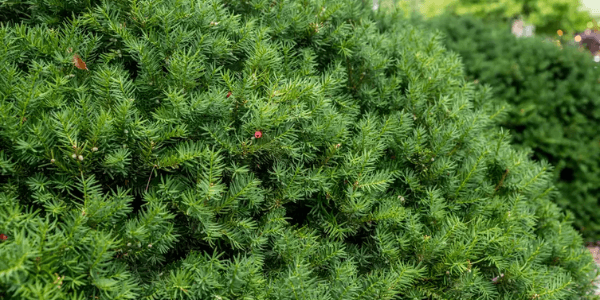
Yew

Yew is commonly found in church yards and even small amounts are fatal. It contains highly toxic alkaloids which affect the heart, causing an irregular heartbeat and rapid death. Yew clippings must be disposed of appropriately and good fencing should prevent cattle from entering church yards and gardens.

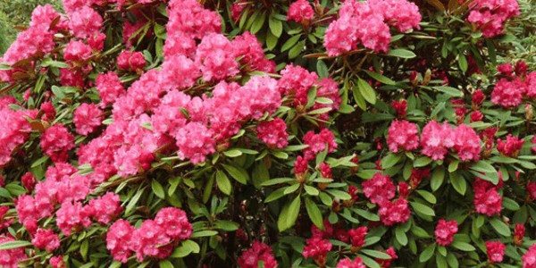
Rhododendron

Rhododendrons contain a poison (Grayanotoxin) that slows the heart rate and lowers blood pressure. Affected livestock are weak, often recumbent, bloated and may vomit, which is unusual for ruminants. Goats in particular my exhibit dramatic projectile vomiting with rhododendron poisoning. Animals show signs of abdominal pain such as groaning and teeth-grinding. Death may be rapid. As a prevention, ensure stock have access to adequate silage or hay at these times to reduce the likelihood of livestock browsing. Pastures bordering woodland should be inspected for rhododendron and fenced accordingly. Ensure clippings are disposed of appropriately.

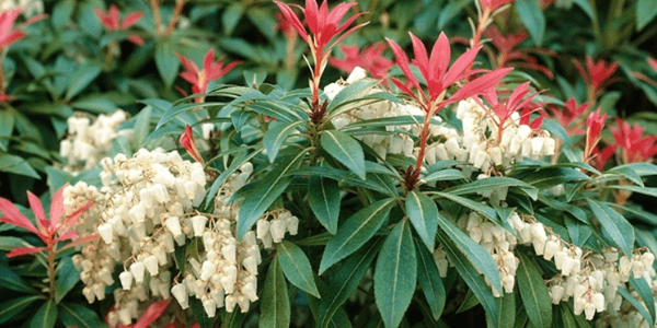
Pieris

Ornamental garden shrub. Most commonly nibbled over a garden fence or ingested when hungry animals break into a garden. This plant contains the same toxin as the Rhododendron (Grayanotoxin) and produces similar gastro-intestinal signs and is often fatal.

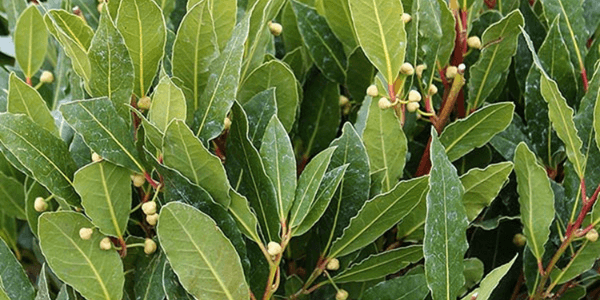
Laurel

Laurel contains cyanide, one of the most rapidly acting toxins affecting cattle. Cyanide prevents haemoglobin in red blood cells from releasing oxygen, effectively starving the tissues of oxygen. Affected animals initially show excitement, increased breathing and heart rate, and increased salivation. The mucous membranes are bright red, eventually turning blue. Muscle spasm, coma and death follow. Laurel should not be planted where animals can reach.

Grass Sickness in horses

Grass Sickness is a disease of horses, ponies and donkeys in which there is damage to parts of the nervous system which control involuntary functions, producing the main symptom of gut paralysis.

Also known as Equine Grass Sickness (EGS) the cause is unknown but the nature of the damage to the nervous system suggests that a type of toxin is involved – potentially botulism neurotoxin acquired from soil.

The toxin may also affect nerves supplying other body systems resulting in other signs of EGS such as droopy eyelids, inability to swallow & muscle tremors to name but a few.

Three forms of the disease have been reported: the acute, subacute and chronic forms. The form a patient succumbs to depends on the extent of nerve damage.

Horses affected by the acute form of the disease present showing signs of colic often indistinguishable from other forms of colic meaning that it may be suspected that the patient has a twisted gut or other form of surgical colic.

As a result, such patients often undergo colic surgery and the diagnosis of EGS is often made presumptively on the surgery table. This form of the disease is 100% fatal.

In horses with subacute or chronic EGS, the time course of the disease is more gradual and these patients may present with a high heart rate, mild episodes of colic, a tucked up appearance, an inability to swallow, drooling saliva, droopy eyelids, muscle tremors and patchy sweating. This form of the disease is also fatal.

Horses with the chronic EGS may survive but require intensive management to maintain hydration and nutritional requirements. The likelihood of survival depends on the extent of nerve damage.

The only way to definitively diagnose EGS is to examine an intestinal biopsy. Surgery is required to obtain a biopsy. Therefore, horses are frequently diagnosed based on the presence of compatible clinical signs.

Vets often perform an eye drop test known as a phenylephrine test. When these drops are applied to one of the patient’s eyes the droopy eyelid appearance improves. Other conditions may also cause droopy eyelids so this test is by no means perfect.

Horses at risk of succumbing to EGS include any horse at grass but the condition is most commonly seen in young animals aged between 2 & 7 years.

Cases have been reported throughout the year but occur most frequently in late spring/early summer. Overweight horses are also at increased risk. Other reported risk factors include recent soil disturbances, overuse of ivermectin based wormers, a recent change in pasture & being at grass 24/7.

Prevention is based on avoiding changes in management, especially in youngstock, at the ‘at risk’ time of year. Soil disturbances should also be kept to a minimum. Ideally, horses should be stabled for at least part of the day and offered hay or haylage. Furthermore, overuse of ivermectin based wormers should be avoided and ideally, a wormer containing an alternative drug should be used prior to turn out. Co-grazing with sheep or cattle may also be protective.

When a case has been diagnosed at a property,it is of paramount importance to stay calm and to avoid any sudden changes in management. In our opinion, in-contact horses should not be moved field as moving pasture is itself a risk factor for

EGS. Furthermore, fields within a 10km radius are theoretically ‘at risk.’ Co-grazing with a patient that has succumbed to EGS may itself be protective suggesting an acquired immunity. We would; however, recommend that young horses are kept off an affected field during future grazing seasons.

A vaccine trial is currently underway which, if licensed, will hopefully provide us with an effective means of preventing EGS in the future.

Tooth decay in horses

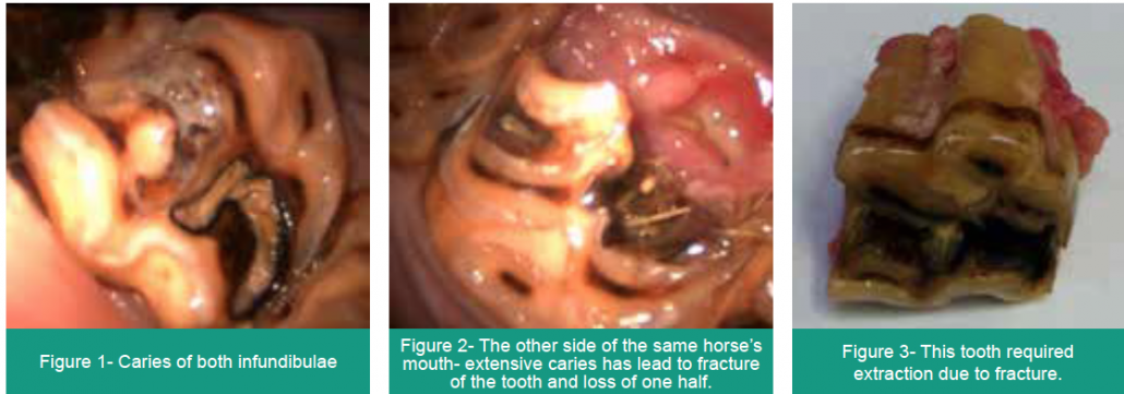
Similar to humans, horses’ teeth suffer from decay, referred to as ‘caries’.

Horses’ upper cheek teeth have infundibulae, cup-shaped invaginations in their grinding surface that are normally filled with a hard material called cementum. Sometimes these infundibulae are incompletely filled during development, leaving a cavity for food to settle in and decay. Over time the decay weakens the structure of the tooth, predisposing it to fracture and, if the decay advances beyond the margins of the infundibulum and into the sensitive pulp system, an apical (root) infection may arise. Both of these situations require extraction of the tooth.

Caries of the infundibulae

How can we prevent this?

Infundibular caries cannot be detected by feel, therefore is it imperative that every horse’s mouth is visually assessed using a headtorch during routine dental examinations to detect these before advanced disease develops.

It is possible to perform cleaning and filling of the cavity with filling material. This is termed an ‘infundibular restoration’. We now have the equipment to perform this at our clinic.

To discuss any of our Advanced Dentistry services, please call Sam Baker at the surgery: 01772 861300

Wild plants poisonous to livestock

During the grazing months it’s important to look out for cases of plant toxicity. Many poisonous plants are bitter and unpalatable while they are growing and will not be eaten by livestock under normal circumstances. However, if grazing is scarce, such as after a period of dry weather, or when pastures are overstocked it can be more likely. Here are a few of the most common wild toxic plants to look out for.

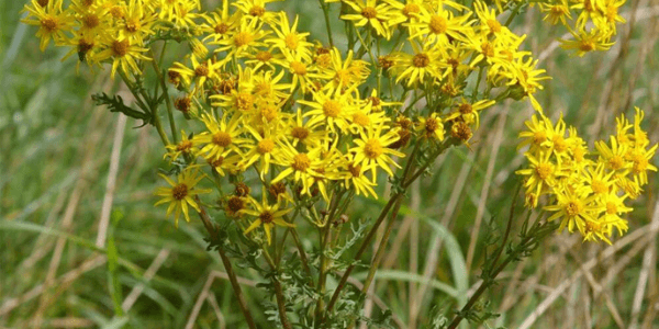
Ragwort

Ragwort poisoning is seen less commonly, as pasture management usually prevents this plant colonising grazing land. Poisoning can occur from ingestion of the live plant or from conserved forages containing Ragwort. Clinical signs of ragwort poisoning include depression, chronic weight loss, diarrhoea, jaundice and accumulation of fluid under the jaw and brisket caused by liver disease. Sheep are much less susceptible to ragwort than cattle. There is no effective treatment once the liver damage has occurred.

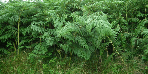
Bracken

Bracken poisoning often occurs when alternative forage is in short supply and animals have little else to eat, additionally some animals develop a taste for the young shoots in the spring. Bracken poisoning requires long-term exposure, over weeks to years. The outcome of ingestion is related to the time period and the quantity ingested. Haematuria (blood in the urine) primarily affects cattle (less commonly sheep) and is often accompanied by anaemia. The toxins cause bone marrow suppression and depletion of white blood cells resulting in immunosuppression. Affected cattle may be weak, exhibit rapid weight loss, high temperature (40°C+), pale mucous membranes, spontaneous bleeding or prolonged bleeding from minor wounds, and blood clots in the faeces. Affected sheep may be blind.

Lower doses of bracken fern ingested over several years are more likely to be carcinogenic and may result in bladder tumours. Poisonings are prevented by not allowing access to bracken, particularly in spring when the young more palatable fronds are emerging. Alternatively, ensure animals have adequate nutrition at all times, so they are not forced to eat bracken as an alternative.

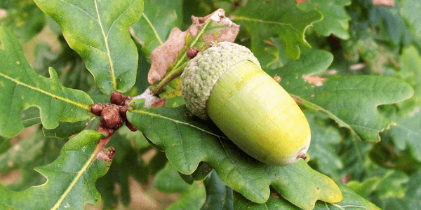
Oak (Acorn)

Acorns contain tannins which cause serious, often fatal, kidney damage. Mass ingestion of acorns may occur following stormy weather. Rumen microbes break down the tannins, producing gallic acid, which causes damage to kidney cells. Animals may show abdominal pain and bloat due to ruminal stasis. They are anorexic, depressed and often recumbent. Constipation may be followed by foetid tarry diarrhoea. Death may occur within hours to several days depending on the number of acorns eaten. It is prudent to fence off oak trees on grazing pastures.

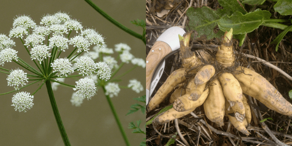
Hemlock water-dropwart

Cattle are particularly at risk of hemlock poisoning after ditches have been cleared out, exposing the most toxic part of the plant, the roots (“dead man’s fingers”). Animals show signs of salivation, dilated pupils, difficulty breathing, collapse and spasmodic convulsions. The majority of affected cattle die.

Bulk milk result round-up

A few weeks ago we collected bulk milk samples from all our dairy farms and screened for the presence of antibodies to IBR, BVD, leptospirosis, liver fluke and Salmonella, depending on their vaccination status. Here’s a summary of the results:

Salmonella dublin

  • Of the 93 farms tested; 25 herds were positive for S. Dublin antibody and 68 herds were negative.
  • This signifies that 27% of the unvaccinated herds we tested had been exposed to S. Dublin!

Leptospirosis

  • 45 herds were unvaccinated for leptospirosis and bulk milk screening revealed 29 tested positive and 16 tested negative.
  • That means that in our sample, 64% of unvaccinated herds had been exposed to leptospirosis.
  • Within the positive herds: 2% had low levels of antibodies, 49% had medium levels and 49% had high levels of antibodies.


BVD

  • Of the 44 herds that were unvaccinated against BVD, 37 farms tested positive for BVD antibodies and 7 were negative.
  • This means that of the unvaccinated farms tested, 84% had been exposed to BVD or had cattle that may have unknown vaccination status.

IBR

  • Of the 110 dairy herds sampled, 28 do not vaccinate against IBR and were tested for antibody status. Of those 28 farms, half tested positive for IBR antibodies and half tested negative.
  • Therefore 50% of the unvaccinated herds that we tested had been exposed to IBR.

Fluke

  • 39 herds opted to screen for liver fluke antibodies.
  • 10 herds screened (25%) were negative, 20 herds (52%) had light infestation, 8 herds (21%) had moderate infestation and 1 herd (2%) had heavy infestation.

Thanks to all farms who took part in this project.

Management of the ‘down cow’

The main reasons for a down cow generally are toxic, metabolic, or traumatic. The longer the cow is down, the worse the prognosis as damage to the leg muscles and nerves begins to occur.

Once the cause of the recumbency has been identified and treatment has been planned, the cow can be moved onto soft, clean bedding – Deep straw, sand (20cm minimum), or ideally out to grass if the weather allows. Have good quality feed and clean fresh water available at all times in a position the cow can easily access. Consider using non-steroidal anti-inflammatories  

A hip hoist can be a useful tool to lift down cows to prevent them from suffering further injury. Ideally use a hip hoist along with a strap under the chest of the cow to take some of the weight. The hip hoist should be firmly attached around the tuber coxae (indicated in the diagram). Lift the cow slowly as vertically as possible with a loader until in a normal weight-bearing position. DO NOT lift the cow up in the air. Allow the cow to stand with the hoist on, if she won’t weight bear then lower her down. Don’t remove the hoist unless confident she is weight-bearing and won’t stagger over.  

All Red Tractor assured farms must now have a written plan in place for the management of down cows. Speak to your vet about making sure yours is upto date.  

Img src: BCVA