CASE STUDY: IT ALWAYS HAPPENS TO A VET’S HORSE!
Jack is a 15-year-old Morgan horse gelding, owned by our vet Roisin, who started with a moderate right hindlimb lameness noticed during schooling exercise. He was admitted to the clinic and had nerve blocks performed which isolated the lameness to the upper cannon region.
X-RAYS & MRI
X-rays and ultrasound scans revealed mild chronic changes to the proximal suspensory ligament but did not fully explain his lameness grade. The proximal suspensory region is a challenging region to image with conventional methods due to superimposition of many anatomical structures over one another, so we elected to put Jack in our MRI (magnetic resonance imaging) machine to give us further information regarding the soft tissues and surroundings bone structures.
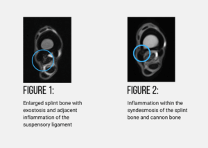
Jack was an MRI star and stood like a rock from start to finish over a few hours in the magnet! The MRI scans echoed ultrasound images, revealing moderate inflammation (desmitis) of the proximal suspensory ligament, but most interestingly revealed a boney protrusion on the inside of Jacks outer splint bone (figure 1.). This was abutting and causing inflammation to the adjacent suspensory ligament. There was also inflammation between the syndesmosis (fibrous join between bones) between this splint and the cannon bone (figure 2.).
RETURNING TO WORK
The scans provided vital information regarding Jack’s likelihood to return to his previous level of exercise. Due to the presence of concurrent pathology to the structures surrounding his suspensory ligament he was unlikely to stay sound for his previous job of hunting and eventing.
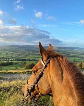
However, Jack was set up on a controlled rehabilitation programme targeted steroid medication and we are happy to say that Jack is sound enjoying a slightly lighter lease of life of pleasure rides, working equitation and hacking, hopefully for many more years to come!